Do you have any questions?
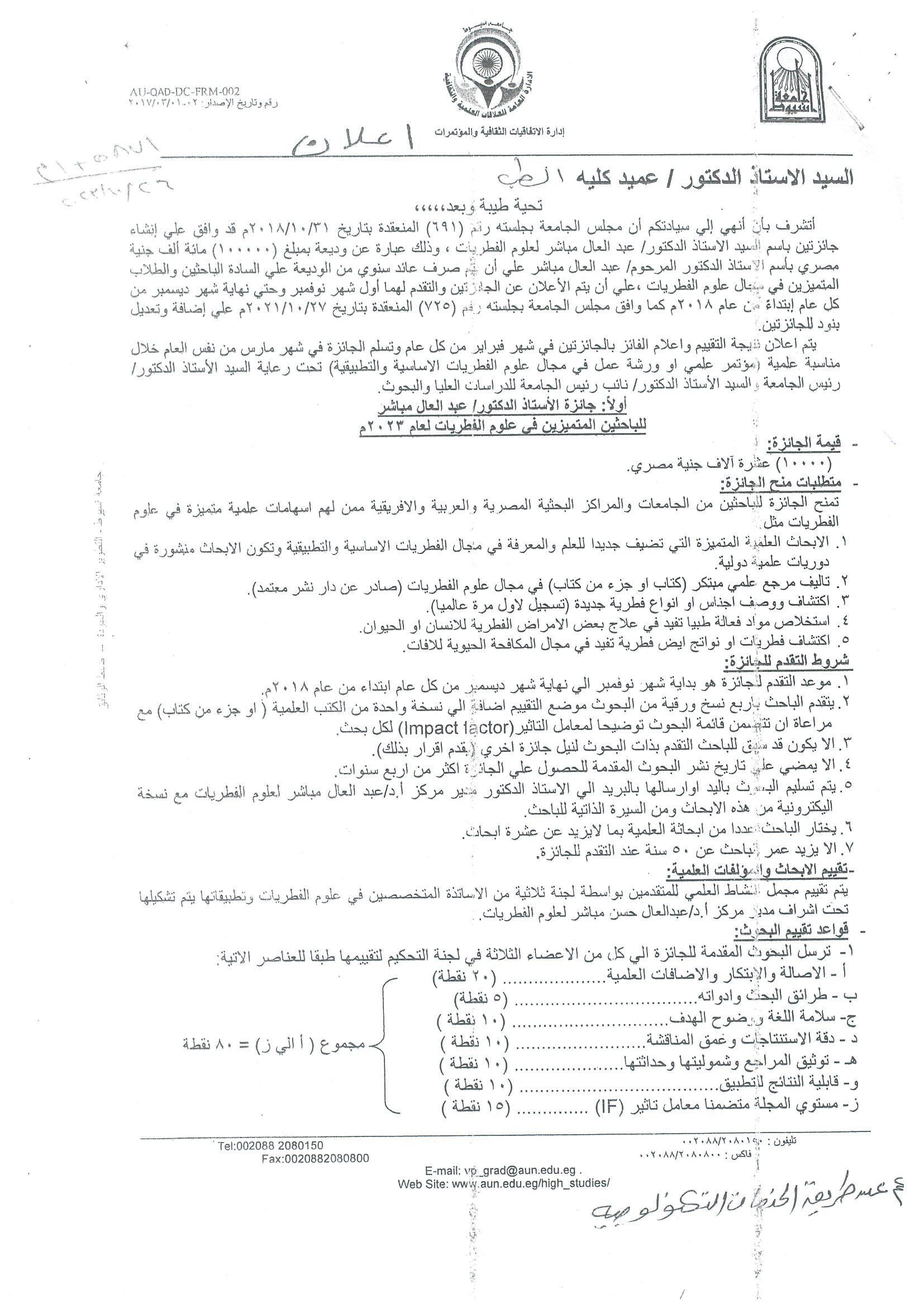
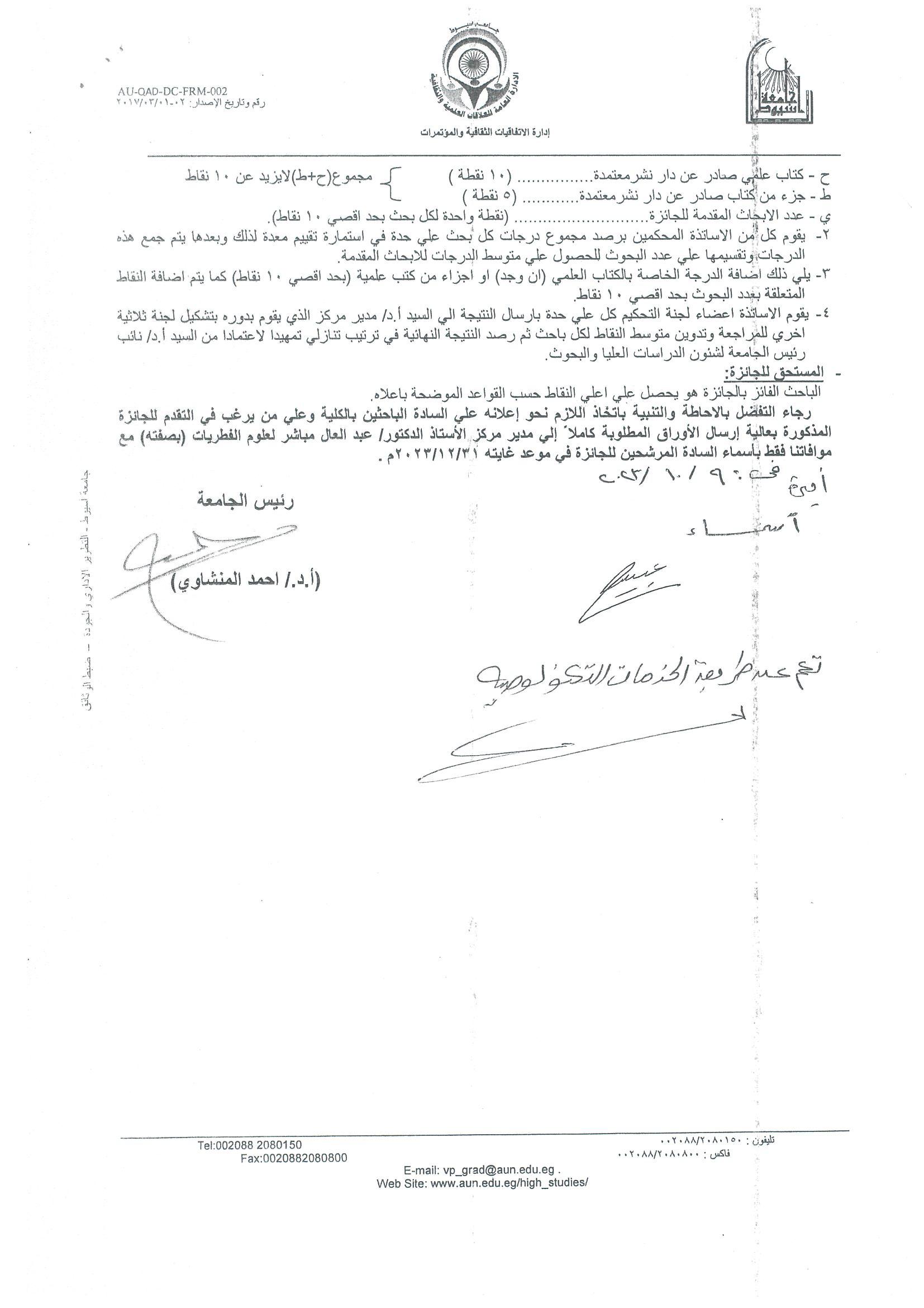
 Announcement regarding Professor Dr. Abdel-Aal Mubasher’s award for mycology
To assess the visual outcome of manual small-incision cataract surgery (MSICS) as well as safety, cost, and time of the procedure.
A retrospective study involving candidates for cataract surgery with baseline-corrected distance visual acuity (CDVA) ≤20/120. Visual acuity (VA) was the primary outcome measure while surgical complications, cost, and time of surgery were the secondary outcome measures. Follow-up visits were scheduled at 1 day, 1 week, 1 month, and at 6 and 12 months following surgery.
The study enrolled 3007 patients with a mean age of 66.45 ± 17.3 years. Out of 3007 patients, 2774 (92.2%) were legally blind before surgery (CDVA <20/200) which was significantly reduced to 55 patients (1.9%) by 1 month following surgery. Uncorrected distance visual acuity was 20/60 or better in 2098 eyes (69.8%) at 1 month, in 2035 eyes (67.7%) at 6 months, and in 2017 eyes (67.1%) at 12 months. The posterior capsular rupture was the most common intraoperative complication. Corneal edema was the most common immediate postoperative complication while the development of posterior capsular opacification was the leading cause for later impaired VA. The mean cost was approximately equivalent to 20 US dollars. The median duration of surgery was 10 min.
MSICS is a safe, cost-effective, and time-saving technique for improving the vision of cataract patients in areas with high cataract surgery volume and limited facilities.
Ocular pain is a common complication following photorefractive keratectomy (PRK). The level of patient satisfaction with current pain control strategies is not high. This study aims to assess the efficacy and safety of a novel regimen of preservative-free oxybuprocaine hydrochloride 0.4% unit-dose eye drops for post-PRK pain control.
In a contralateral eye study, 144 eyes of 72 patients who underwent bilateral transepithelial PRK (TransPRK) were stratified into experimental and control groups. The experimental group received preservative-free oxybuprocaine hydrochloride 0.4% unit-dose eye drops five times daily postoperatively until complete epithelial healing, while the control group received sodium hyaluronate 0.2% instead. The main outcome measures were pain scores assessed by the verbal rating scale and visual analogue scale (VRS, VAS), the corneal epithelial defect (CED) area, epithelial healing duration evaluated by slit-lamp biomicroscopy and anterior segment optical coherence tomography (AS-OCT), and endothelial cell density (ECD) measured before and 1 month after surgery.
Pain scores assessed by VRS and VAS were significantly lower in the experimental group 8 h after surgery, and 1, 2, and 3 days postoperatively (P < 0.001). The mean CED area showed no significant differences between the two groups at different follow-ups (P value > 0.05). The corneal epithelial healing had a mean duration of 3.32 ± 0.47 days in both studied groups and was parallel in both eyes of each patient. In each group, 49 eyes (68%) and 72 eyes (100%) had a fully epithelialized surface on the third and fourth postoperative days, respectively. No significant changes were observed in the mean ECD 1 month following surgery in both groups (P value > 0.05).
Preservative-free oxybuprocaine hydrochloride 0.4% unit-dose eye drops are effective and safe in controlling early postoperative pain following TransPRK. The availability of the single-dose unit preparation can overcome the problem of topical anesthetic abuse.
Objective:
To assess and compare postlaser in situ keratomileusis (LASIK) dry eye after LASIK with planned thin flaps created by a femtosecond laser (FS) and mechanical microkeratome (MK).
Methods:
Patients were stratified according to the flap creation technique into FS and MK groups with planned 100 μm flap thickness in all eyes. Dry eye parameters including tear film break-up time (TBUT), Schirmer I test, ocular surface disease index (OSDI), and lower tear meniscus height and area (tear meniscus height [TMH] and tear meniscus area [TMA]) were assessed before surgery and at 3 and 6 months after surgery.
Results:
The study included 102 eyes of 55 patients (52 eyes underwent FS-LASIK/50 eyes underwent MK-LASIK). The preoperative characteristics including age, gender, and spherical equivalents were similar in both groups (P> 0.05). The difference in postoperative flap thickness was statistically …
Conventional mechanical or alcohol-assisted photorefractive keratectomy (PRK) techniques for correction of hyperopia and hyperopic astigmatism were associated with inconsistent results. The aim of this study is to evaluate the 12-month visual and refractive outcomes of the relatively new single-step transepithelial photorefractive keratectomy (TE-PRK) for moderate hyperopia and hyperopic astigmatism.
Methods
This is a prospective interventional study. Forty-eight eyes of 30 patients with moderate hyperopia or hyperopic astigmatism with a cycloplegic spherical equivalent refraction (SEQ) between 2.0 and 4.5 diopters (D) underwent single-step StreamLight® TE-PRK using EX500 excimer laser (Alcon Laboratories, USA). The main outcome measures were recorded at 6 and 12 months postoperatively including assessment of logarithm of the minimum angle resolution (logMAR) uncorrected and …
Objectives
To assess and compare the six-month outcome of the two-step transepithelial phototherapeutic keratectomy- photorefractive keratectomy (PTK-PRK) and the single-step transepithelial PRK for myopia and myopic astigmatism.
Methods
A prospective randomized study. The study enrolled 100 eyes of 50 patients with mild to moderate myopia or myopic astigmatism stratified into two groups, PTK-PRK (n = 50 eyes) and single step PRK (n = 50 eyes). Primary outcome measures were visual acuity and manifest refraction. Secondary outcome measures were epithelial healing duration, post-PRK pain scores and 3-month postoperative haze grading.
Results
Preoperative characteristics were similar in both groups (p value > 0.05). The mean uncorrected distance visual acuity (UDVA) at 1 week, 1 month, 3 and 6 months was significantly better in the single-step PRK group than in the two-step PTK-PRK group …
Background
Corneal collagen cross-linking (CXL) is a procedure utilized for halting keratoconus progression with different approved protocols. The current study aimed to assess the corneal endothelial changes following the relatively new accelerated pulsed high-fluence protocol of epithelium-off corneal cross-linking for the treatment of mild to moderate keratoconus.
Methods
This prospective case series study enrolled 45 eyes of 27 patients with mild to moderate progressive keratoconus who underwent accelerated pulsed high-fluence CXL (pl-ACXL, 30 mW/ cm2 UVA at 365 nm wavelength, 8 min pulsed mode 1 s on / 1 s off with a total energy of 7.2 J/ cm2). The main outcome measures were corneal endothelial changes assessed by specular microscopy at 3 and 6 months postoperatively including endothelial cell density (ECD), coefficient of variation, percentage of hexagonal cells, average, minimum and …