 Do you have any questions?
 Seminar by Dr. Ashraf Mohamed Rashwan - Specialist in the Department of Ophthalmology - Assiut University Hospital
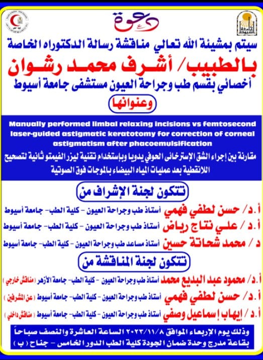
Seminar by Dr. Ashraf Mohamed Rashwan - Specialist in the Department of Ophthalmology - Assiut University Hospital
Seminar by Dr. Ashraf Mohamed Rashwan - Specialist in the Department of Ophthalmology - Assiut University Hospital
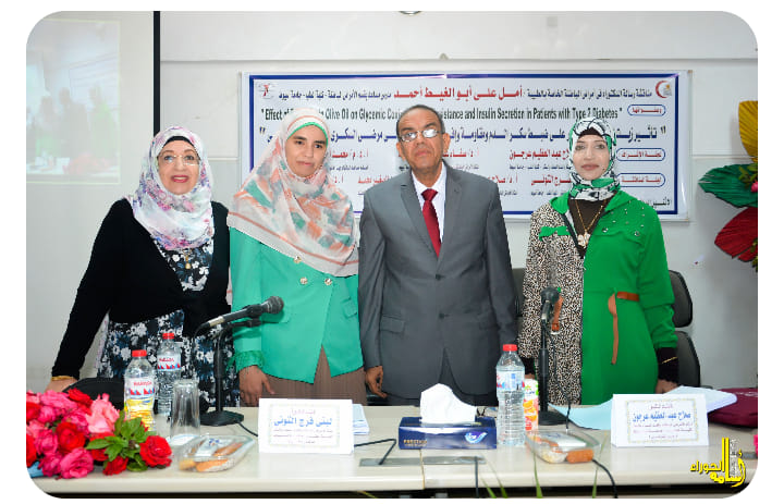
Objectives We aimed to study the prevalence of each type of pulmonary hypertension (PHT) (precapillary, post capillary and combined) in end stage renal disease patients (ESRD) under haemodialysis (HD). We also studied the correlation between the systolic pulmonary artery pressure (SPAP) and different patient clinical and laboratory variables. Methods This cross-sectional study was conducted on 106 HD patients. Demographic and clinical data, blood samples for laboratory variables of the studied patients were all collected. A standard echocardiography was done. Pulmonary function test was performed with standard spirometry. Results The total prevalence of PHT in our study population was 77.3%. The prevalence of isolated post capillary PHT, pre capillary PHT and combined pre and post capillary PHT was (32.07%, 23.58% 21.69%) respectively. The prevalence of “unexplained” PHT was 0.9%. There were significant positive correlation between SPAP and smoking index (r=0.427, P=0.035), duration of dialysis (r=0.416, P=0.046), ferrittin level (r=0.312, P=0.048), cardiac output (CO) (r=0.683, P=0.000) and AV fistula flow (r=0.529, P=0.018), while there were significant negative correlation between SPAP and hemoglobin (HB) level (r=−0.598, P=0.010) and serum iron (r=−0.572, P=0.049). Multivariate analysis showed significant association with smoking index (P=0.01), duration of dialysis (P=0.032), HB level (P=0.042) and CO (P=0.000) after adjusting other factors. Conclusions In our study there was a high prevalence of PHT in HD patients with a very low prevalence of the unexplained types of PHT. Smoking index, duration of dialysis, HB level and CO were independent risk factors of increased SPAP.
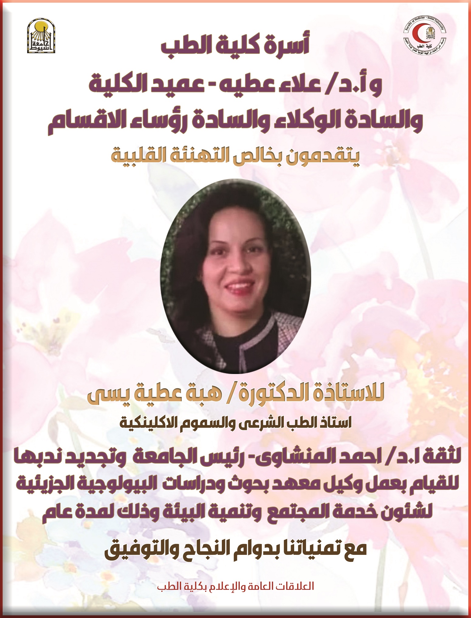
Congratulations to Mrs. Prof. Dr. Heba Attia Yassa - for renewing her secondment to carry out the work of Undersecretary of the Institute for Molecular Biological Research and Studies
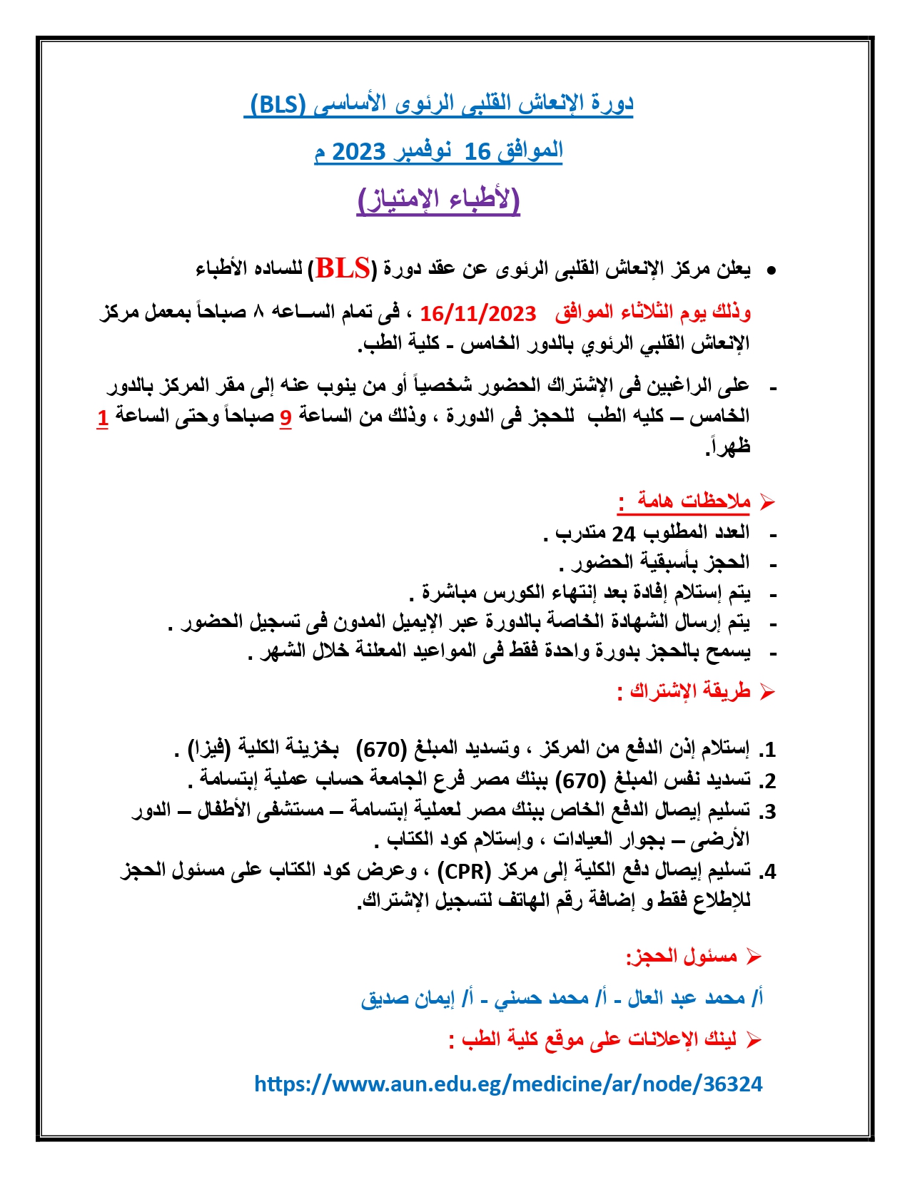
Announcement of the Cardiopulmonary Resuscitation Center (BLS) course, November 16, 2023

Announcement of the Cardiopulmonary Resuscitation Center (BLS) course, November 13, 2023
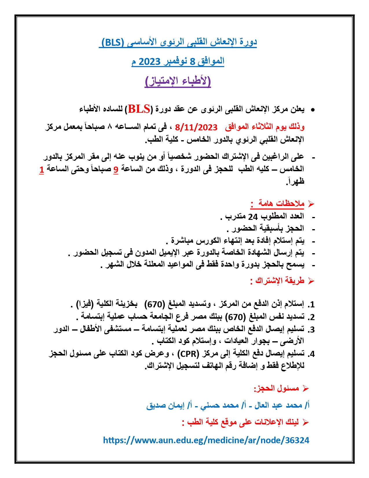
Announcement of the Cardiopulmonary Resuscitation Center (BLS) course, November 8, 2023
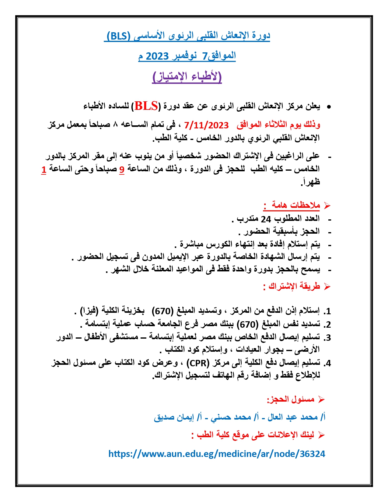
Announcement of the Cardiopulmonary Resuscitation Center (BLS) course, November 7, 2023
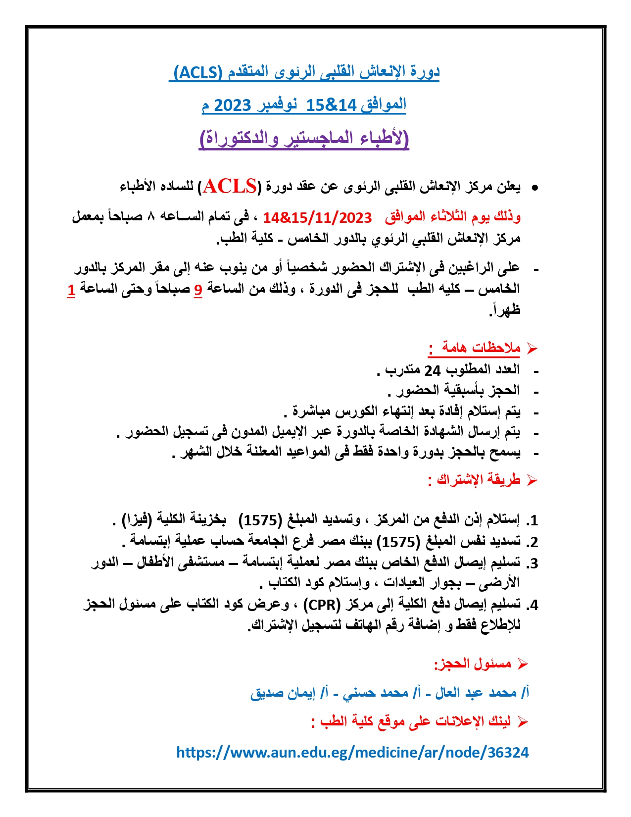
Announcement of the Cardiopulmonary Resuscitation Center (ACLS) course, November 2023
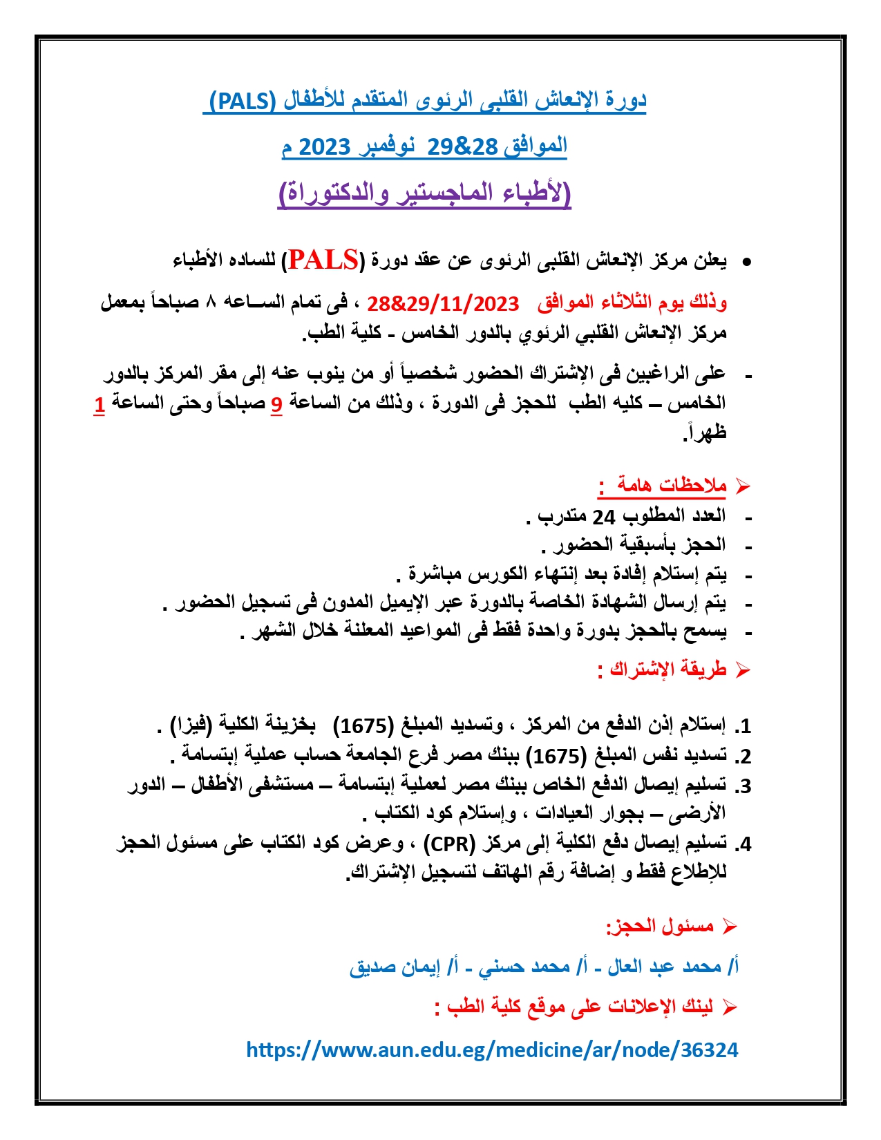
Announcement of the Cardiopulmonary Resuscitation Center (PALS) course, November 2023