 Do you have any questions?
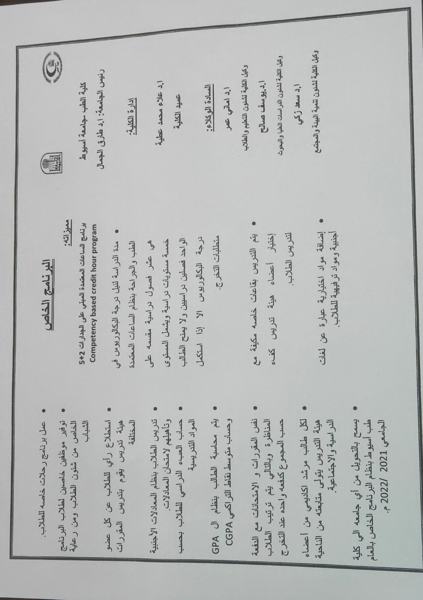
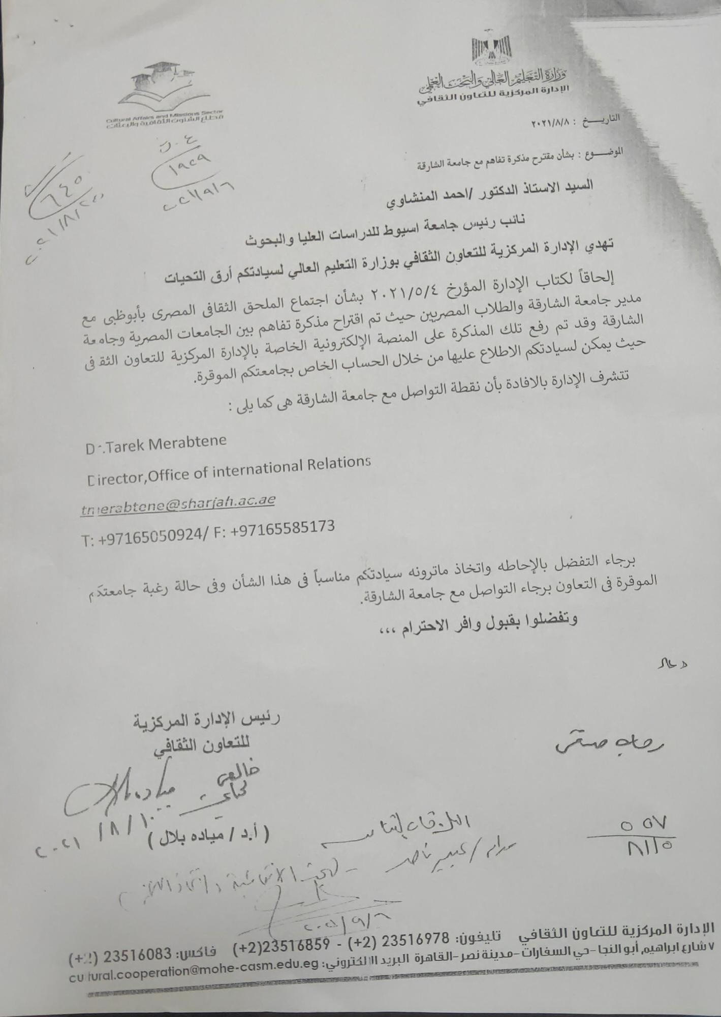
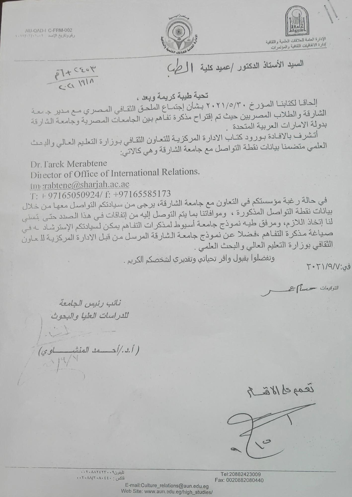
A memorandum of understanding was proposed between Egyptian universities and the University of Sharjah
Impact of sleep deprivation and sleep recovery on reproductive hormones and testicular oxidative stress in adult male rats
Research Abstract
Objectives: sleep deprivation is a significant problem among adult men. It is considered to be a risk factor that contributes to several disease. It has been proposed that reactive oxygen species and the resulting oxidative stress may be responsible for some of the effects of sleep deprivation. The present study was performed to determine the impact of sleep deprivation for different periods on serum testosterone, luteinizing hormone, corticosterone and whether sleep deprivation causes oxidative stress indicated by measuring malondialdehyde (MDA) level in testicular tissue as a direct evidence of cellular damage and measuring glutathione (GSH) in testicular tissue to determine possibility of reversible nature of oxidative stress by scavenger antioxidant system. We studied also if sleep recovery after sleep loss could relieve these effects or not. Material and methods: 42 adult male albino rats aged 12 weeks weighing about 200-250 gm were used in this study. They were divided into seven groups, six animals in each group, a control group and six experimental groups. Three experimental groups were used as sleep deprivation (SD) groups and another three experimental groups were used as sleep recovery (SR) groups. The SR groups were also sleep deprived and then returned to home-cages and were allowed to undisturbed and spontaneous sleep. Group I: served as a control group. Group II: rats were subjected to sleep deprivation for one day. Group III: rats were subjected to sleep deprivation for three days. Group IV: rats were subjected to sleep deprivation for five days. Group V: rats were subjected to sleep deprivation for three days followed by a period of sleep recovery for one day. Group VI: rats were subjected to sleep deprivation for three days followed by a period of sleep recovery for three days. Group VII: rats were subjected to sleep deprivation for three days followed by a period of sleep recovery for five days. After each planned SD and SR period, blood samples were collected for hormonal assay. The rats were decapitated and the testes were dissected out and used for the study of malondialdehyde and glutathione. The parameters were measured then analyzed by using Student's t-test. Results: serum testosterone level and luteinizing hormone (LH) showed significant decrease after three days of deprivation. Serum corticosterone level increased significantly from the first day of deprivation in comparison with the control group. After five days of sleep recovery, serum testosterone level and corticosterone returned to the level of the control group. Serum LH level improved after three days of sleep recovery. Sleep deprivation increased the testicular tissue MDA significantly and GSH was significantly decreased after three day of sleep deprivation when comparing with the control. Sleep recovery decreased testicular tissue MDA significantly and significant increase in GSH after the fifth day as non-significant change noticed on comparing their levels on that day with the control. Conclusions: The present study demonstrated the sleep deprivation effects on testosterone, luteinizing hormone, corticosterone levels in serum, malondialdehyde and glutathione in testicular tissue of rat. Sleep recovery was associated with restoration of the serum hormone levels. Also with sleep recovery MDA level was decreased and GSH content was improved in testicular tissue.
Research Date
Research Department
Research Journal
Al-Azhar Assuit Medical Journal
Research Member
Functional and Structural Study on the Effect of Curcumin on Folic Acid-Induced Acute Kidney Injury in Albino Rats
Research Abstract
|
Objective. Systemic administration of folic acid (FA) in rats was used for studying the pathogenesis associated with acute renal damage. However, the mechanism by which FA induces renal damage remains poorly understood. Up to our knowledge, no effective preventive or therapeutic drugs have been developed to protect against acute kidney injury. Curcumin (CUR) is commonly used worldwide as a spice and has been demonstrated to possess various biological activities. The present study was planned to investigate the effect of folic acid administration on renal function, inflammatory cytokines and associated histological changes in renal tissue. In addition, we examined the possible protective effect of curcumin on a rat model of folic acid (FA)-induced acute kidney injury (AKI). Methods. Rats were divided into 3 groups; (FA) folic acid treated group rats were exposed to FA (250 mg/kg) i.p. injection as a single dose. (FA+CUR) folic acid plus curcumin treatment group rats were given curcumin (200 mg/kg) administered by gavage daily for 11 days prior to folic acid (250 mg/kg) i.p injection and the last dose of curcumin was given one day after folic acid injection. Control group are given distilled water by gavage daily for 12 days and saline i.p. as a single dose on the 11th day. Animals were scarified one day following i.p. injection in all groups. Deterioration of kidney function was detected by blood urea and creatinine levels. Inflammatory response was monitored with blood levels of interleukin-6 (IL-6), interleukin-10 (IL-10), and tumor necrosis factor (TNF-α). Results. We found that FA treatment significantly raised blood urea, creatinine, IL-6, IL-10, TNF-α levels and caused marked structural changes of the kidney. CUR treatment for 12 days significantly reduced blood urea, IL-6, IL-10, TNF- α, and protected partially against renal structural damage. Conclusion. These findings suggest that curcumin is a promising protective agent against AKI induced by FA. |
Research Date
Research Department
Research Journal
Bull. of Egyp. Soc. Physiol. Sci.
Mechanism of action of oxytocin as cardioprotection in rat myocardial infarction
Research Abstract
Oxytocin (OT) is well known for its role in reproduction. However, evidence has emerged suggesting a role in cardiovascular system but less is known about the role of this hormone in the injured heart. We elucidate oxytocin cardioprotective effects against myocardial infarction (MI). Male rats were divided into six groups: control without surgery, sham without occlusion, MI, OT pretreated then MI, combined OT and L-NAME (NO synthase inhibitor) then MI and combined OT and indomethacin (cyclooxygenase blocker) then MI. Twenty-four hours post-MI induction, hemodynamics parameters, inflammation markers, oxidative stress markers, apoptotic gene expression, brain naturitic peptide (BNP), and histopathological assessment were carried out. When compared to MI model group, OT significantly reduced LVEDP and increased LVSP and ± dP/dt.Also, it significantly decreased serum levels of BNP, TNF-α, IL-6, and TBARS with an increase in the activities of SOD and GPx. Furthermore, BAX and p53 mRNA were decreased. Interestingly, no significant improvement in any of the markers was detectable when we administrated OT with L-NAME. While the same results observed when we treated the rat with OT and indomethacin. We conclude that OT protected against the sequelae of myocardial infarction. These findings provide new insight into therapeutic strategies for myocardial infarction.
Research Date
Research Department
Research Journal
IOSR Journal of Dental and Medical Sciences
Gastroprotective effect of flavonoid quercetin and coenzyme Q10 in indomethacin-induced gastric ulcers in normal and diabetic rats
Research Abstract
Various studies have indicated that peptic ulcers occurring during the course of diabetic state are more severe and often associated with complications such as gastrointestinal bleeding. This study is an attempt to understand the pathogenesis of indomethacin-induced gastric ulcers occurring during the diabetic state using suitable markers and its amelioration by quercetin and coenzyme Q10 (CoQ10). In this study, diabetic rats showed an increase in the gastric mucosal levels of Molandialdehyde (MDA), inducible nitric oxide synthase (iNOS), interleukin-6 (IL-6), tumor necrosis factor (TNF-α), BAX and p53 and a decrease in the activities of superoxide dismutase (SOD) as compared to normal control (non-diabetic) rats. There was an increase in gastric ulcer index and gastric ulcer lesions in diabetic gastric mucosa when compared to the normal control group. Pre-treatment with quercetin and\or CoQ10 to normal groups or diabetic groups which treated by indomethacin caused a significant decrease in gastric ulcer index, MDA, iNOS, IL-6, TNF-α, BAX and p53 with concomitant increase in SOD activity when compared with normal and diabetic rats treated with indomethacin alone. So quercetin and CoQ10 are effective in protection against indomethacin-induced gastric ulcers in normal and diabetic rats. Our findings could bring new hope for a novel modality of gastric ulcer treatment.
Research Date
Research Department
Research Journal
IOSR Journal of Dental and Medical Sciences
Neuroprotective Effect of Resveratrol Against Brain Ischemia Reperfusion Injury In Rats Entails Reduction of DJ-1 protein expression and Activation of PI3K/Akt/GSK3b survival Pathway
Research Abstract
In the current study, we aimed to investigate the mechanistic role of DJ-1/PI3K/Akt survival pathway in ischemia/reperfusion (I/R) induced cerebral damage and to investigate if the Resveratrol (RES) mediates its ischemic neuroprotection through this pathway. RES administration to sham rats, boosted GSH level and SOD activity and downregulated iNOS expression without affecting redox levels of DJ-1 forms nor components of PI3K/Akt pathway including PTEN, p-Akt or p/p-GSK3b. However, RES pre-administration to I/R rats, reduced infarction area, oxidative stress, inflammation and apoptosis. Concomitantly, RES ameliorated the decreased levels of oxidized forms of DJ-1 and enhancing its reduction, increased the nuclear protein expression of Nfr-2 and led to activation of PI3K/Akt survival pathway. In conclusion, overoxidation of DJ-1 is a major factor that contributes to post I/R cerebral damage andits reduction by RES could explain the neuroprotection offered by RSE.
Research Date
Research Department
Research Journal
Archives of Physiology And Biochemistry
Vitamin D Aggravates the Metabolic Side Effects of Olanzapine in Female Rats
Research Abstract
Objective: Atypical antipsychotics represented a major advance in the treatment of schizophrenia and minimizing the extrapyramidal side effects. However, the use of atypical antipsychotics have been linked to weight gain, hyperglycemia, metabolic syndrome and risk of liver affection. Previous studies proposed that vitamin D deficiency may contribute to the development of insulin resistance, metabolic syndrome and more recently fatty liver. The goal of this study wasto investigate the role of vitamin D in protection against the metabolic and hepatic side effects of olanzapine.
Methods: Eighteen female albino rats received treatment by gavage for 5 weeks and divided into; C group: received (0.5ml/day) of normal saline and olive oil (0.2ml) twice weekly, (O) group: received olanzapine (2mg/ kg/day) and olive oil (0.2ml) twice weekly, and (O+D) group: received olanzapine (2mg/kg/day) and vitamin D3 (vit.D3) (125mcg/ Kg) orally by gavage twice weekly. Plasma levels of lipid panel, liver enzymes, glucose, insulin, Interleukin-6 (IL-6), Interleukin-10 (IL-10), and Tumor necrosis-alpha (TNF-α)were determined. H & E staining of liver tissue were performed to assess the effects of vit.D3 treatment on olanzapine-induced histopathology.
Results: Co-administration of vit.D3 caused significant elevation of IL-6, IL-10, HDL/LDL and mild improvement of liver histopathology. However, it caused further elevation of triglycerides (TGs), total cholesterol (TC), very low density lipoproteins (VLDL), TNF-α, liver enzymes, plasma, bilirubin, and impaired glucose tolerance. Insignificant difference in weight gain and abdominal fat was found.
Conclusion: These results suggest that olanzapine-induced disturbed lipid profile and hepatic steatosis is independent of weight gain.Moreover, this study provides an evidence for adverse effects of vitamin D supplementation especially in patients treated with olanzapine.
Research Date
Research Department
Research Journal
IOSR Journal of Dental and Medical Sciences
Rutin hydrate inhibits apoptosis in the brains of cadmium chloride-treated rats via preserving the mitochondrial integrity and inhibiting endoplasmic reticulum stress
Research Abstract
Recent evidence has suggested that cadmium (Cd) ions-induced neurotoxicity is associated with increased oxidative stress and mitochondrial-dependent and endoplasmic reticulum (ER) stress-induced apoptosis. This study aimed to investigate if rutin hydrate (RH), a well-reported neuroprotective and an antioxidant flavonoid, can ameliorate cadmium chloride (CdCl2)-induced neurotoxicity by inhibiting the resultant ER stress. Rats were divided into 4 groups (n = 16/group) of control, control + RH (100 mg/kg), CdCl2 (5 mg/kg), and CdCl2 + RH. All treatments were administered orally for 30 days, on daily basis. Brain homogenates from CdCl2-treated rats showed increased oxidative stress and induced activation of ER stress characterized by increasing mRNA and protein levels of GRP78, ATF-6, CHOP and Xbp-1 and protein levels of p-elF2α, p-JNK1/2 and cleaved caspase-12. Also, CdCl2 significantly reduced Bcl-2, enhanced Bax translocation to the mitochondrial membrane, increased cytoplasmic levels of cytochrome-C and caspase-3, and reduced mitochondrial membrane potential (Δψm) (increased Vmax and reduced time to Vmax). In contrast, RH significantly enhanced levels GSH and activities of SOD, GSH-Px, decreased levels of MDA and inhibited mitochondrial permeability transition pore (mtPTP) in the brains of both control and CdCl2-treated rats. Interestingly, in brain homogenates of CdCl2-treated rats only, RH reduced all markers of ER stress, increased Bcl-2, reduced mitochondrial Bax translocation and improved mitochondrial coupling. It also reduced cytosolic levels of cytochrome-C, cleaved caspase-3, and cleaved caspase-12. Overall, these findings support the efficiency of RH to inhibit ER stress in brains CdCl2-treated rats which is added to its existing mechanisms of neuroprotection.
Research Date
Research Department
Research Journal
Neurological research
Resveratrol protects against hepatic insulin resistance in a rat's model of non-alcoholic fatty liver disease by down-regulation of GPAT-1 and DGAT2 expression and inhibition of PKC membranous translocation.
Research Abstract
Non-alcoholic fatty liver disease (NAFLD) is associated with hepatic insulin resistance (IR). Resveratrol (RES) a potent hypolipidemic dietary polyphenol has been identified for its ability to prevent hepatic steatosis and hepatic IR in high-fat diet (HFD)-fed murine models of NAFLD. In the present study, we have carried an in vivo animal experiment to identify a novel mechanism for RES protective action. Sub-chronic (45 days) RES pretreatment in 3 days HFD-fed adult Wistar rats prevented early hepatic IR through inhibiting PKC/JNK activation; decreasing p-IRS (Ser307) and increasing p-IRS (Tyr612), p-Akt (Ser473) and p-GSK3(Ser9). These effects of RES were associated with reduced expression of acyl-CoA:glycerol-sn-3-phosphate acyltransferase (GPAT-1) and diacylglycerol:acyl-CoA acyltransferase (DGAT2), two critical enzymes in the glycerol-3-phosphate pathway for de novo triglycerides synthesis. These data indicate that RES protects against NAFLD, initially, by inhibiting the early development of hepatic IR.
Research Date
Research Department
Research Journal
Clinical and Experimental Pharmacology and Physiology