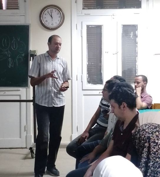
Toxic cardiomyopathies were a potentially fatal adverse effect of anthracycline therapy.
This study was conducted to demonstrate the pathogenetic, morphologic, and toxicologic effects of doxorubicin on the heart and to investigate how the MAPK /TNF-α pathway can be modulated to improve doxorubicin-Induced cardiac lesions using bone marrow-derived mesenchymal stem cells (BM-MSCs) and olive leaf extract (OLE).
During the study, 40 adult male rats were used. Ten were used to donate MSCs, and the other 30 were split into 5 equal groups: Group I was the negative control, Group II obtained oral OLE, Group III obtained an intraperitoneal cumulative dose of DOX (12 mg/kg) in 6 equal doses of 2 mg/kg every 48 h for 12 days, Group IV obtained intraperitoneal DOX and oral OLE at the same time, and Group V obtained intraperitoneal DOX and BM-MSCs through the tail vein at the same time for 12 days. Four weeks after their last dose of DOX, the rats were euthanized. By checking the bioinformatic databases, a molecularly targeted path was selected. Then the histological, immunohistochemistry, and gene expression of ERK, JNK, NF-κB, IL-6, and TNF-α were done.
Myocardial immunohistochemistry revealed severe fibrosis, cell degeneration, increased vimentin, and decreased CD-31 expression in the DOX-treated group, along with a marked shift in morphometric measurements, a disordered ultrastructure, and overexpression of inflammatory genes (ERK, NF-κB, IL-6, and TNF-α), oxidative stress markers, and cardiac biomarkers. Both groups IV and V displayed reduced cardiac fibrosis or inflammation, restoration of the microstructure and ultrastructure of the myocardium, downregulation of inflammatory genes, markers of oxidative stress, and cardiac biomarkers, a notable decline in vimentin, and an uptick in CD-31 expression. In contrast to group IV, group V showed a considerable beneficial effect.
Both OLE and BM-MSCs showed an ameliorating effect in rat models of DOX-induced cardiotoxicity, with BM-MSCs showing a greater influence than OLE.
Background
Bisphenol-A (BPA) has a well-proven deleterious effect on the hypothalamicpituitary- gonadal axis.
Objectives
The current study investigated the therapeutic potentials of mesenchymal stem cells (MSCs) in a murine model of BPA-induced ovarian damage.
Methods
Fifty adult female rats were divided into: Group 1; control group, Group IIa, IIb: rats were given oral gavage of BPA (25 and 50 mg/Kg body weight respectively) on a daily basis for 15 days, and Group IIIa, IIIb; rats were intravenously treated with of MSCs (106 cells) after receiving the last dose of BPA as in group II. Plasma and ovarian tissue levels of Malondialdehyde (MDA) and gonadal axis hormones were assessed. Apoptosis was evaluated by TUNNEL assay and by apoptosis markers (FAS, FASL, Caspase 3, SLTM). A histological examination of ovarian tissue was also conducted.
Results
BPA resulted in a significant elevation in plasma …