The Computer Center announces the seventh session for postgraduate doctors... online... and in person
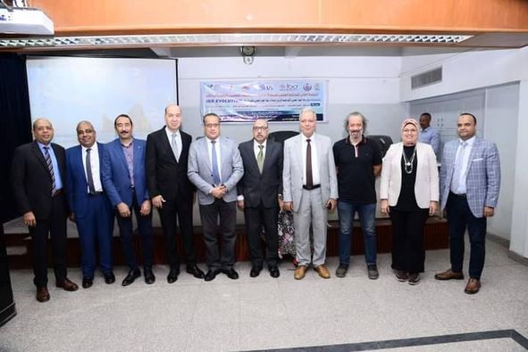
Activities of the first session of the IBD EVOLUTION scientific program on developments in the diagnosis and treatment of colon and intestinal immune diseases
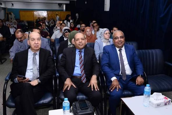
الاستاذ الدكتور علاء عطية عميد كلية ورئيس مجلس إدارة المستشفيات الجامعية
وبحضور الأستاذ الدكتور محمد عبد الرحمن، وكيل الكلية لشئون التعليم والطلاب
الأستاذ الدكتور/ هدى مخلوف، وكيل الكلية لشئون خدمة المجتمع وتنمية البيئة.
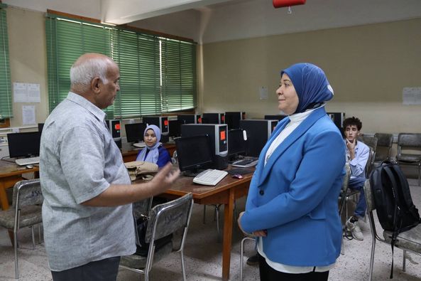
الأستاذ الدكتور/ فرج محمد مفتاح المشرف على الحاسب الآلي.
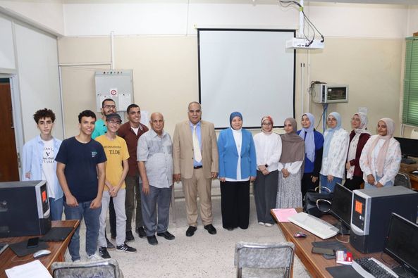
عقد يوم الأربعاء ٢٥ سبتمبر اجتماع بمركز الحاسب الآلى لفريق البحث العلمى لطلاب طب أسيوط (١٣ طالب)،، لمتابعة نتائج تدريب الطلاب علي البحث العلمى والاحصاء الطبى، وذلك من خلال دورتين سبق تنظيمها من خلال وحدة الحاسب الآلى بكلية الطب فى حوالى (٢٢ ساعة) تدريب .
وقد قام الطلاب بتقسيم أنفسهم إلى خمسة مجموعات بحثية لكل منهم بروتوكول بحثى.
وبعد اطلاع السادة الوكلاء علي الأبحاث المطروحة، قاموا بالنقاش مع الطلاب لتحديد التخصص المناسب لكل بحث ، وتحديد عدد من المشرفين لمتابعة البرتوكول مع الطلاب.
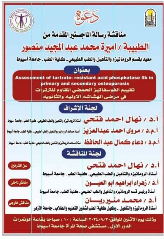
Seminar by Dr. Amira Mohamed Abdel Majeed Mansour - Teaching Assistant in the Department of Rheumatology, Rehabilitation and Physical Medicine - Faculty of Medicine - Assiut University
Seminar by Dr. Amira Mohamed Abdel Majeed Mansour - Teaching Assistant in the Department of Rheumatology, Rehabilitation and Physical Medicine - Faculty of Medicine - Assiut University
The Training Unit has the honor of conducting a cultural course and symposium on the use of artificial intelligence in the field of obstetrics and gynecology
Congratulations to Dr. Hisham Al-Bassit - Pediatric Orthopedic Teacher
 Do you have any questions?