Do you have any questions?
 Prohibition of attendance at work Only after submitting a proof of obtaining the Corona virus vaccine
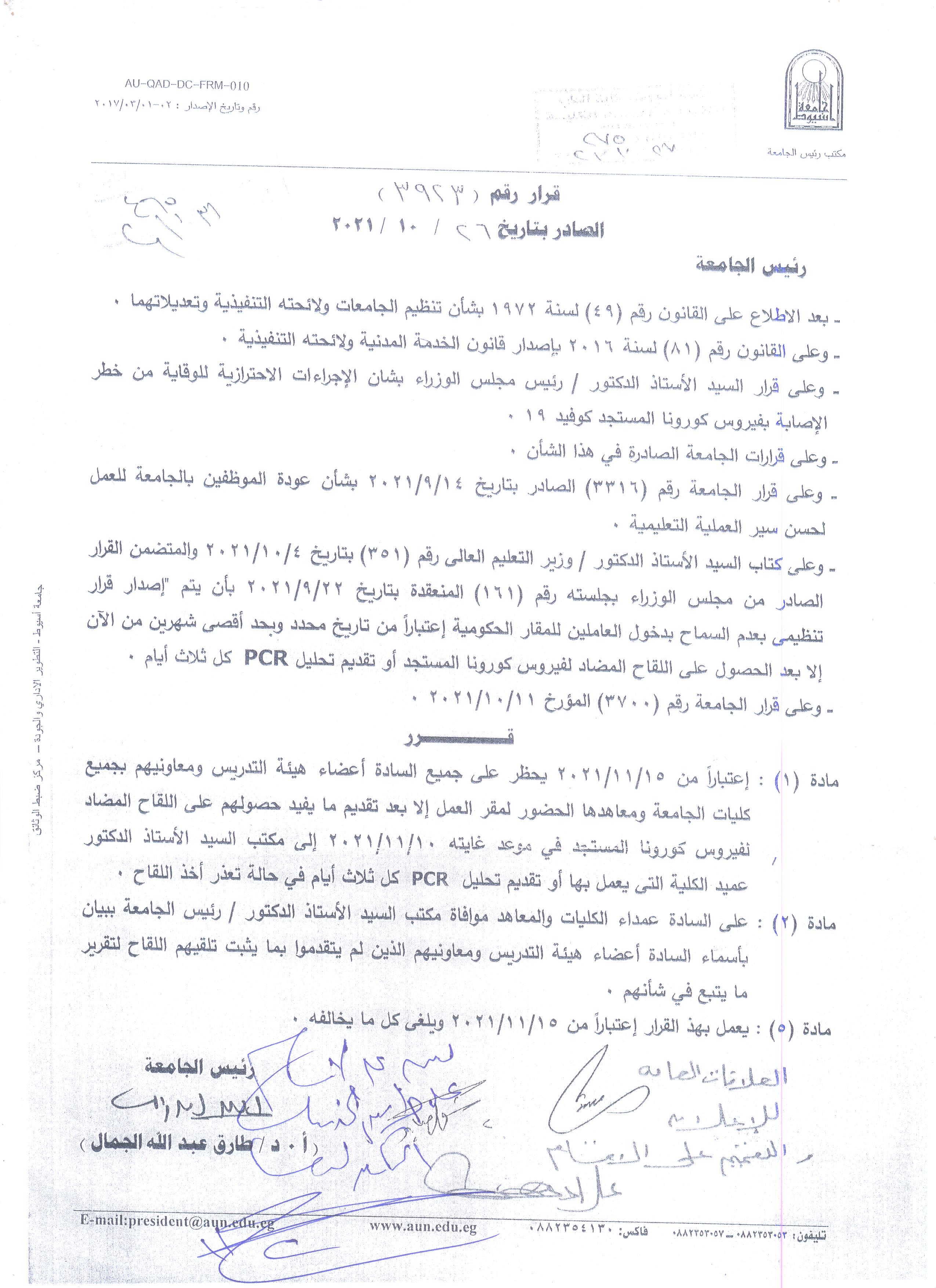
Prohibition of attendance at work
Only after submitting a proof of obtaining the Corona virus vaccine
Prohibition of attendance at work
Only after submitting a proof of obtaining the Corona virus vaccine
Eimeria sp. is one of the most important parasites that cause very high economic loss in
poultry farms in Egypt. It causes a disease called coccidiosis. This study aimed to detect the
prevalence of Eimeria species in chicken and detection of its pathological effect within the
intestinal mucosa. The present study investigate the prevalence of Eimeria sp. in chicken
through fecal examination and the diagnosis was based on direct fecal sample examination
(unstained wet mount technique) and concentration techniques, followed by sporulation of
unsporulated oocyst for identification of Eimeria spp. and finally studying the pathological
effect of this parasite in the intestinal mucosa of infected chicken. The total prevalence rate of
Eimeria spp. was (66%). The incidence rate in Broiler chickens was (70%) and in Balady was
(58%). The highest percent of infection was at the age of (15-30) day (54.3% in Broiler and
72.4% for Balady), and the disease was more prevalent in winter than in summer. The species
that were detected are E.acovullina (the highest prevalence rate) followed by E.tenella,
E.necatrix followed by E.mitis (lowest prevelance rate). These results indicate that the
coccidiosis is a serious parasitic disease that effect on the poultry production in Egypt and
control measures should be put in consideration to overcome this disease.
Keyswords: Eimeria - prevalence- coccidiosis- oocyst
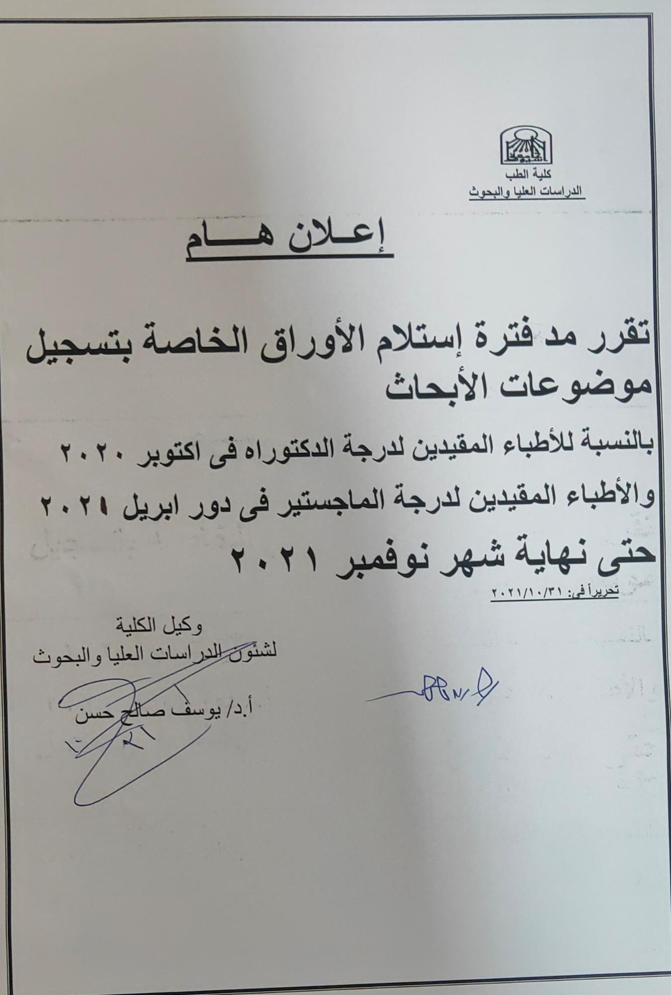
 مد فترة إستلام الأوراق الخاصة بتسجيل موضوعات الأبحاث بالنسبة للأطباء المقيدين لدرجة الدكتوراة والماجستير 2021

 Applications for the Research Grants Program have begun
Gastrointestinal parasites have a direct effect on the health of equines. This study
was conducted to determine the prevalence and significance of gastrointestinal
parasites of naturally infected horses in Luxor Governorate, Egypt. A total of 100
horse fecal samples were collected during the period from March - 2020 to February
– 2021 from different ages, sexes and stables. All of these samples were examined
by different fecal examination techniques including (direct wet mount,
sedimentation and flotation techniques). The present study investigate that the total
prevalence of infected horses was 15%. There is a relationship between the GIT
infection and the age of the horse. The highest percent of infection was recorded by
Strongylus vulgaris 9 % followed by Parascaris equorum 5%, while the percent of
infection by Balantidium coli was 1% in infected horses. It consequence that horses
are highly susceptible to Strongylus irrespective of gender and age or even
deworming. Control measures should be put in consideration to totally overcome the
parasitic infection.
Keywords: Gastrointestinal parasites, Parascaris equorum, Strongylus vulgaris,
Balantidium coli.
Cystic echinococcosis has been considered one of the major parasitic zoonoses which is associated with severe economic losses. The present study was undertaken to investigate the occurrence, organ distribution, cyst fertility, and viability of cystic echinococcosis in slaughtered camels and cattle from various abattoirs in Assiut Governorate, Egypt. The work also involved morphological, morphometric, and molecular identification of the parasite. The occurrence of hydatid cysts was investigated in total number of 100 lungs of camels and 574 liver and lungs of cattle admitted to three slaughterhouses at Assiut Governorate, Egypt. Moreover, several individual variable factors, including organ involvement, age, sex, and hydatid cyst characteristics, were studied to identify their possible association with the occurrence of the disease. Genomic DNA was extracted from the hydatid cysts, followed by molecular identification of the parasite through amplification of ribosomal DNA internal transcribed spacer (ITS) regions. Hydatid cysts were found in 6 camels (6%) out of 100 inspected camels, while 5 hydatid cysts (0.87%) were detected in a total number of 574 cattle examined. The parasite was detected exclusively in lungs of camels, while lungs were the main organ infected by the parasite in cattle and one hydatid cyst was found in the liver (0.17%). In camel, 66.7, 16.65, and 16.65%of detected cysts were fertile, sterile, and calcified, respectively, while in cattle, these percentages were 60, 20, and 20%, respectively. None of the studied variable factors were significantly associated with the occurrence of the disease in camels, with the exception that all cysts were found in the lung. Conversely, we found a significant association (P < 0.05) between the age and sex of the slaughtered cattle and the occurrence of hydatid cysts. In this respect, the rate of infection was higher in female cattle and those cattle more than 5 years (P < 0.05). The morphological, morphometric, and molecular studies confirmed the presence of the parasite. Taken together, our results concluded that camels and cattle play a potential role in maintaining the transmission cycle of this zoonotic parasite.
afaf a abdel wareth m s hasanein assiut med j vol 15 no 6 november 1991