
Press
?
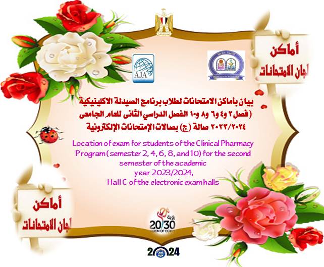
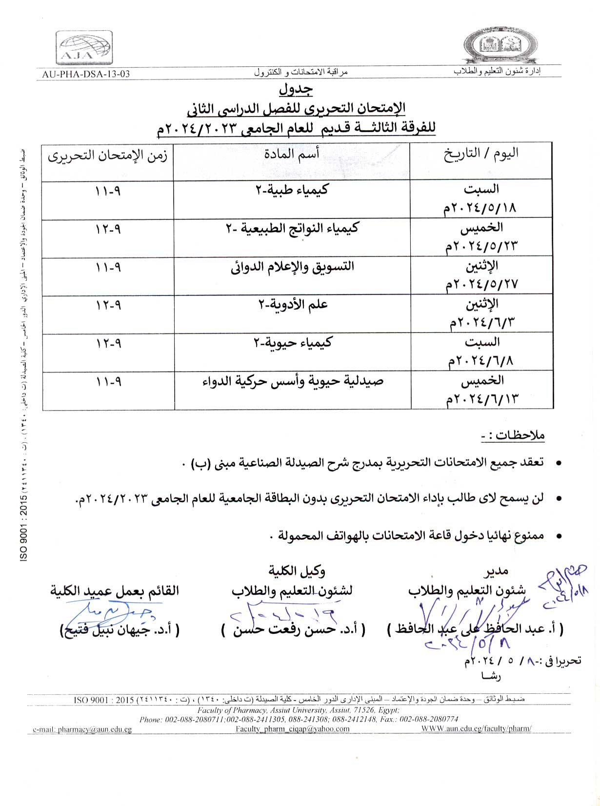
Statement of exam locations for students (first, second, third, fourth and fifth grade)

 Do you have any questions? (088) 2080369 - 2345622 Pharmacy_QAAU@pharm.aun.edu.eg
Wound healing is one of the most challenging medical circumstances for patients. Pathogens can infect wounds, resulting in tissue damage, inflammation, and disruption of the healing process. Simvastatin was investigated recently, as a wound healing agent that may supersede the present therapies for wounds. Our goal in this paper is to focus on formulation of simvastatin cubosomes for topical delivery, as a potential approach to improve simvastatin skin permeation. By this technique its wound healing effect could be improved. Cubosomes were prepared using the top-down method and the prepared cubosomes were characterized by several techniques. The most optimal simvastatin cubosomal formulation was then included in a cubogel dosage form using different gelling agents. The results showed that the average particle size of the prepared cubosomes was 113.90 ± 0.58 nm, the entrapment efficiency was 93.95 ± 0.49% and a sustained simvastatin release was achieved. The optimized formula of simvastatin cubogel displayed pseudoplastic rheological behavior. This same formula achieved enhancement in drug permeation through excised rat skin compared to free simvastatin hydrogel with flux values of 46.18 ± 2.12 mcg cm−2 h−1 and 25.92 ± 3.45 mcg cm−2 h−1 respectively. Based on the in-vivo rat studies results, this study proved a promising potential of simvastatin cubosomes as wound healing remedy.

God willing, a meeting of the committee for community and environmental development will hold on Monday, May 13, 2024, at ten o’clock in the morning.
In the office of Vice Dean for Community Services and Environmental Development Affairs.

God willing, the laboratories and scientific equipment committee will hold its meeting on Monday, May 13, 2024, at eleven o’clock in the morning.
in the office of Vice Dean for Community Services and Environmental Development Affairs.

This current study reports, for the first time, on the potent cytotoxicity of (Z)-3-hexenyl-β-D-glucopyranoside, as well as
its cellular and molecular apoptotic mechanisms against Panc1 cancer cells. The cytotoxicity of three compounds, namely
(Z)-3-hexenyl-β-D-glucopyranoside (1), gallic acid (2), and pyrogallol (3), which were isolated from C. rotang leaf, was
investigated against certain cancer and normal cells using the MTT assay. The cellular apoptotic activity and Panc1 cell cycle
impact of compound (1) were examined through flow cytometry analysis and Annexin V-FITC cellular apoptotic assays.
Additionally, RT-PCR was employed to evaluate the effect of compound (1) on the Panc1 apoptotic genes Casp3 and Bax,
as well as the antiapoptotic gene Bcl-2. (Z)-3-hexenyl-β-D-glucopyranoside demonstrated the highest cytotoxic activity
against Panc1 cancer cells, with an IC50
value of 7.6 μM. In comparison, gallic acid exhibited an IC50
value of 21.8 μM, and
pyrogallol showed an IC50
value of 198.2 μM. However, (Z)-3-hexenyl-β-D-glucopyranoside displayed minimal or no significant
cytotoxic activity against HepG2 and MCF7 cancer cells as well as WI-38 normal cells, with IC50
values of 45.8 μM,
108.7 μM, and 194. μM, respectively. (Z)-3-hexenyl-β-D-glucopyranoside (10 μM) was demonstrated to induce cellular
apoptosis and cell growth arrest at the S phase of the cell cycle in Panc1 cells. These findings were supported by RT-PCR
analysis, which revealed the upregulation of apoptotic genes (Casp3 and Bax) and the downregulation of the antiapoptotic
gene Bcl-2. This study emphasizes the significant cellular potency of (Z)-3-hexenyl-β-D-glucopyranoside in specifically
inducing cytotoxicity in Panc1 cells.
Abstract
Background Calamus rotang L. (CR) is an Indian shrub. The leaves and other organs of the plant are traditionally used
in India for treatment of various diseases. The in vitro antioxidant property of the leaves extract was previously established.
Thus, the current study aimed to evaluate the antioxidant and hepatoprotective effects of CR ethyl acetate
extract at a dose of 350 mg/kg on CCl4
induced hepatotoxic rats through different mechanisms.
Methods Histopathological examination of the treated rats’ group in comparison with positive and negative controls
were performed. Quantitative measuring of the proinflammatory cytokines (TNF α), inflammatory regulators
(Arginase, PPAR α) and the antiapoptotic protein Bcl-2 in comparison with positive and negative control groups was
achieved using immunohistochemical examination. HPLC profiling of the polyphenol contents and molecular docking
of the identified compounds against BH3 proapoptotic protein were correspondingly studied to evaluate the
potential antiapoptotic property.
Results The CR extract greatly protects the liver tissue through the suppression of TNF α, arginase and PPAR α
induced by CCl4
as well as its enhancement of the antiapoptotic Bcl-2 protein. Fourteen polyphenols of different
classes were identified in CR extract and tested via molecular docking for their potential antiapoptotic activities
against BH3 protein. Naringin, rutin, 7-hydroxy flavone, and ellagic acid compounds exhibit the highest affinity and
potential inhibition of pro-apoptotic protein BH3 via molecular docking study.
Conclusions The ethyl acetate fraction of the leaves of C. rotang is rich in polyphenols that exhibited potent hepatoprotective
effect on CCl4
induced hepatotoxic rats through its antioxidant, anti-inflammatory, anti-steatosis and
antiapoptotic properties

God willing, the Clinical Pharmacy Department Council will hold its regular monthly meeting number (101) on Thursday, May 9, 2024 at at 12 :00 AM.
in the meetings Hall of the Department - 5th floor (Building A)
In the department council meeting room