 Do you have any questions?
 Announcement of a workshop entitled (Kalamak Aya initiative) to teach sign language
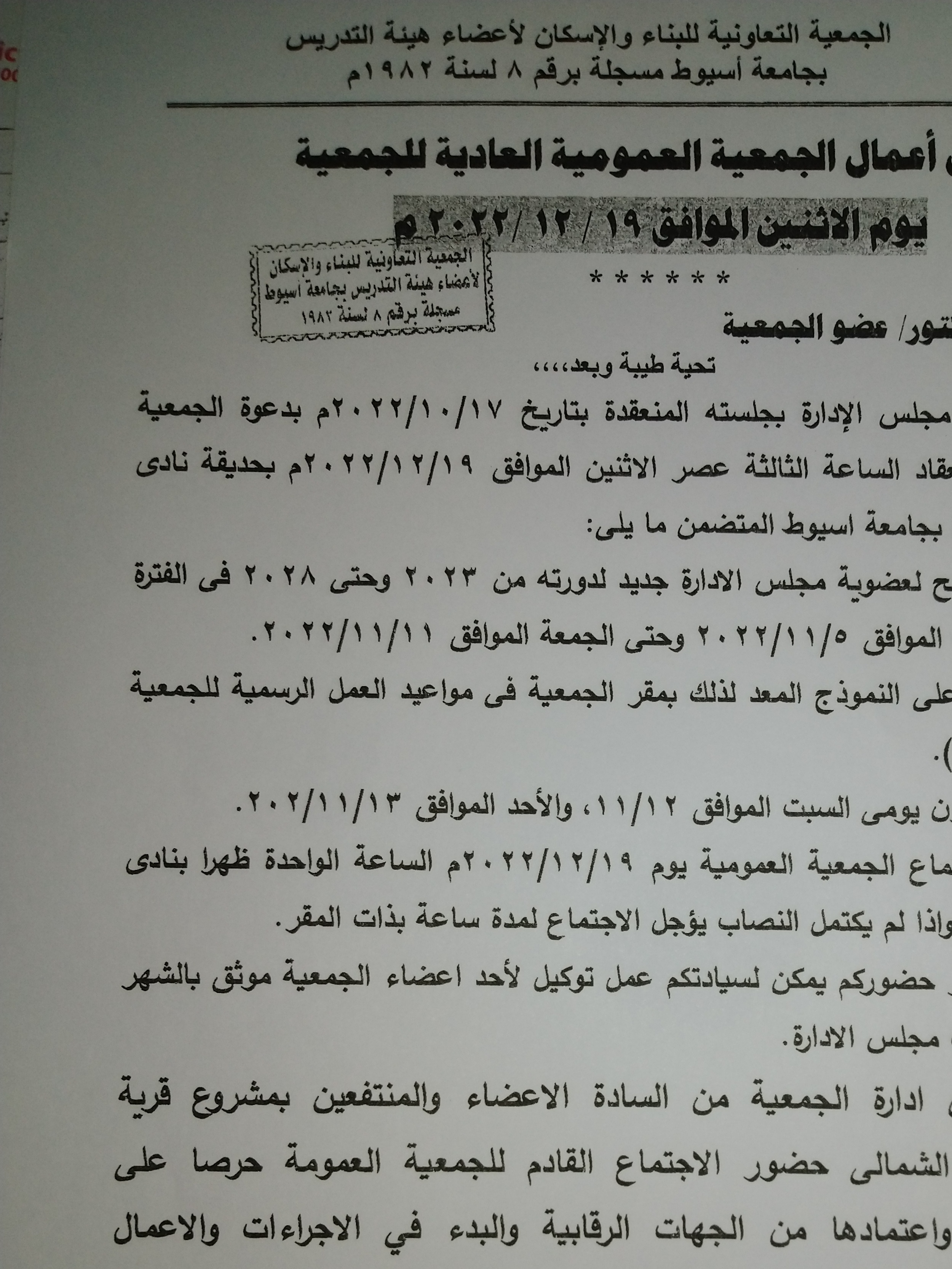
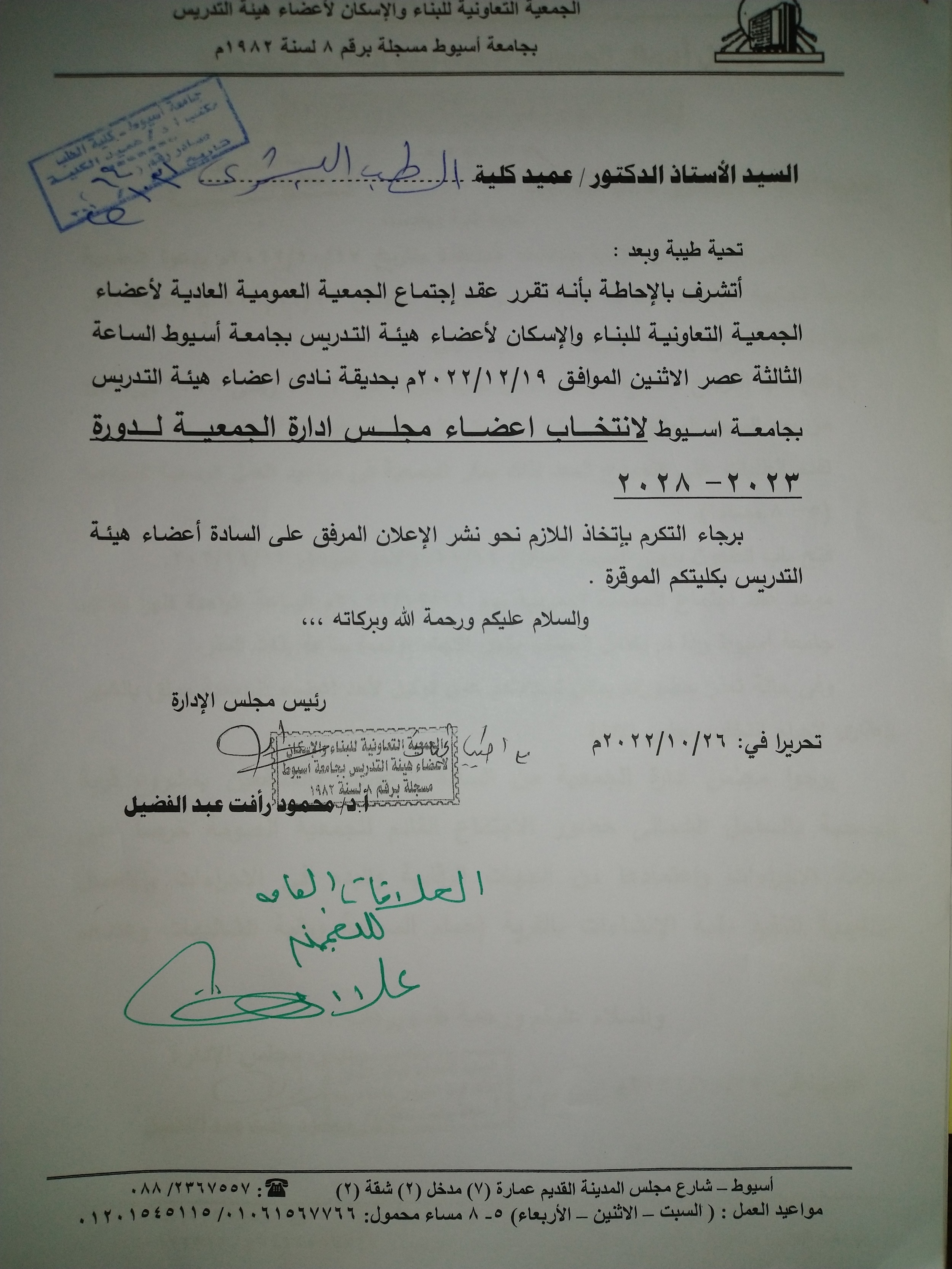
The meeting of the general assembly of the members of the cooperative society for construction and housing for faculty members at Assiut University
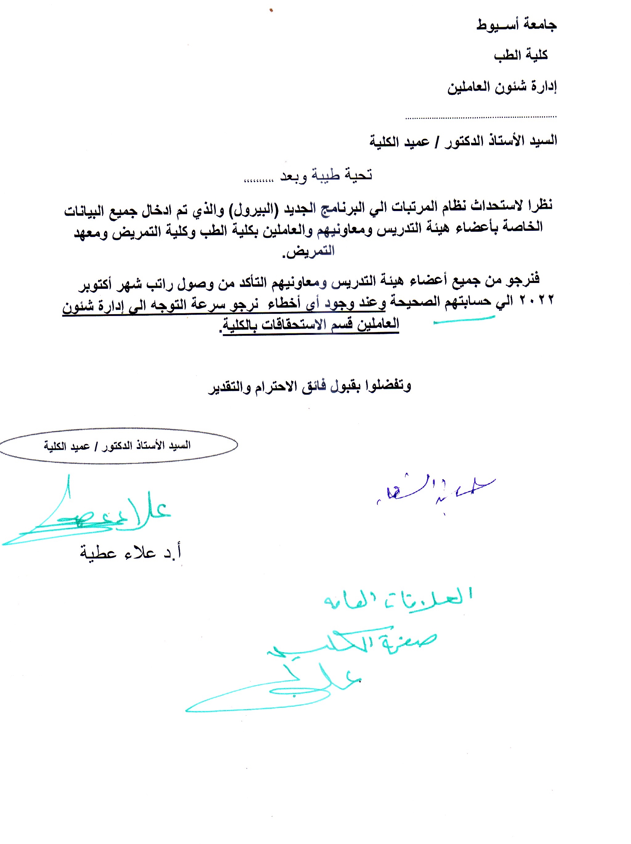
Important Announcement Regarding - Salaries
Enhancement of β-Glucan biological activity using a modified acid-base extraction method from Saccharomyces cerevisia
Research Abstract
Beta glucan (β-glucan) has promising bioactive properties. Consequently, the use of β-glucan as a food additive is favored with the dual-purpose potential of increasing the fiber content of food products and enhancing their health properties. Our aim was to evaluate the biological activity of β-glucan (antimicrobial, antitoxic, immunostimulatory, and anticancer) extracted from Saccharomyces cerevisiae using a modified acid-base extraction method. The results demonstrated that a modified acid-base extraction method gives a higher biological efficacy of β-glucan than in the water extraction method. Using 0.5 mg dry weight of acid-base extracted β-glucan (AB extracted) not only succeeded in removing 100% of aflatoxins, but also had a promising antimicrobial activity against multidrug-resistant bacteria, fungi, and yeast, with minimum inhibitory concentrations (MIC) of 0.39 and 0.19 mg/mL in the case of resistant Staphylococcus aureus (MRSA) and Pseudomonas aeruginosa, respectively. In addition, AB extract exhibited a positive immunomodulatory effect, mediated through the high induction of TNFα, IL-6, IFN-γ, and IL-2. Moreover, AB extract showed a greater anticancer effect against A549, MDA-MB-232, and HepG-2 cells compared to WI-38 cells, at high concentrations. By studying the cell death mechanism using flow-cytometry, AB extract was shown to induce apoptotic cell death at higher concentrations, as in the case of MDA-MB-231 and HePG-2 cells. In conclusion, the use of a modified AB for β-glucan from Saccharomyces cerevisiae exerted a promising antimicrobial,
Research Date
Research Journal
Molecules
Research Member
Research Year
2021
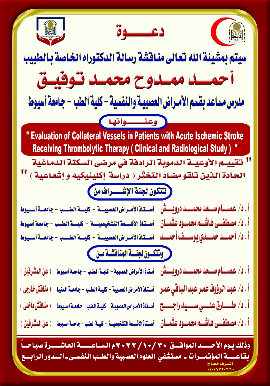
Doctor's Seminar - Ahmed Mamdouh Mohamed Tawfik - Assistant Lecturer - Neurological and Psychiatric Diseases
Pharmaceutical Seminar - Nada Helmy Abdel Tawab
Role of Copper-Albumin Complex in Treatment of Gastric Ulcer in Rats
Research Abstract
The main goal of this submitted work was to investigate the effect of copper-albumin complex on serum and mucosal oxidative stress for gastric ulcer treatment. Forty seven 8-week-old rats were classified into five groups as follow: G1 (control, n=7), G2 (Experimental control, n=10) was left without treatment, G3 (Mucogel group, n=10), G4 (Mucogel-plus group, n=10) and G5 (Omeprazole group, n=10). Treatment has been started six hour after Induction of Ulcers and continued till the 6th day. Ulcer index were reduced more than 30% in G3 and about 90% in G4 more than that of G5 in comparison to G2. The immunostained fundic tissue showed a marked reduction of iNOS activity in G4 more than in G3 and G5. Nitric oxide (NO) was slightly reduced in serum of G3 but it was somewhat at the level of reference control by treatment in G4 and G5 which recorded the strongest reduction of mucosal NO level followed by G4 then G3 compared to G1. Moreover, serum and mucosal total peroxide and oxidative stress index (OSI) were highly elevated in G2 while G4 recorded the strongest reduction followed by G5 then G3 compared to G1. On the other hand, Serum and mucosal superoxide dismutase (SOD) activity showed a significant reduction in G2 and G3 from that of G1 but significantly increased in G4 followed by G5. We also noted that total antioxidant activity (TAO) levels in both Serum and mucosa was almost normalized in G4 followed by G5 then the serum TAO in G3 but mucosal TAO level was non-significantly different comparing G2 and G3. Thus, co treatment of copper-albumin complex seems to be useful for gastric ulcer treatment by decreasing oxidative stress.
Research Date
Research Department
Research Journal
Journal of Applied Sciences Research
Research Member
Research Pages
5789-5798
Research Vol
8 (12)
Research Year
2012
Copper one (I)-Nicotinate Promotes Gene Expression of CYPs that Produce M1 and Q1 in Aflatoxicosed Rats
Research Abstract
Abstract: Aflatoxin-contaminated food poses a serious risk to both human
and animal health. Copper (1)-nicotinate (Cu+-nicotinate) complex
potentiated the prophylactic effect against chronic aflatoxicosis in the
experimental animals through the synthesis of less toxic metabolites M1
and Q1 which are easily excreted in urine. To investigate the action of the
safety Cu+-nicotinate complex on gene expression of Cytochrome 450
(CYP450) system, the liver tissue samples of orally administered rats for 3
weeks with aflatoxin B1 (AFB1; 30 μg/kg body weight), with and without
association of the copper complex (400 μg/kg body weight) were assayed
for their gene expression of CYP450 families including 1A2, 3A2 and
2C11 as well as histopathological examination of the hepatic tissue samples
was performed. The obtained data denoted that the Cu+-nicotinate complex
significantly reduced gene expression of CYP2C11 and CYP3A2 that
enhancing the most toxic epoxide metabolite. On the contrary, this complex
enhanced the expression of CYP1A2 that synthesize the less toxic
metabolite M1 and Q1. The histopathological examination mostly
confirmed such observation as the signs of aflatoxicosis were absent in
Cu+-nicotinate-treated group. Consequently, it could be predicted that the
Cu+-nicotinate complex may be medically used as an inhibitory food
additive agent against exposure of aflatoxicosis in the intact animals since
the complex contains the copper and nicotinic acid, the two daily required
biochemical elements for sustaining live in healthy conditions.
Research Date
Research Department
Research Journal
American Journal of Biochemistry and Biotechnology
Research Member
Research Pages
40-50
Research Publisher
Science Publications
Research Vol
16 (1)
Research Year
2020
IRON-KINETICS IN ALBINO RATS
Research Abstract
A mathematical model of iron kinetics derived by POLLYCOVE and MORTIMER (1961) was successfully used on normal adult rats. R.B.C. iron turnover number, mean delay of iron in red marrow for haemoglobinization, and the R.B.C. life span were determined. Comp < /strong>lete iron exchange between the main plasma iron pool, and the storage compart ments was detected. The findings elucidated the comp < /strong>lex equilibrium between the plasma iron and the various iron pools, namely the erythrocytic pool, itself com prising almost equal labile and fixed compartments and the storage pool. The equil ibrium shows that the iron actually deposited in the storage pool is little (97 Pg Fe/day) comp aired to the erythrocytic iron turnover (410 Pg Fe/day).
Research Date
Research Department
Research Journal
Assiut Veterinary Medical Journal
Research Member
Research Pages
45-50
Research Vol
8 (15)
Research Year
1981