 Do you have any questions? (088) 2080369 - 2345622 Pharmacy_QAAU@pharm.aun.edu.eg
Important announcement for students of the fifth year of the Bachelor of Pharmacy program

Important announcement for fifth-year students in the Bachelor of Pharmacy program (Clinical Pharmacy)
Important announcement for fifth-year students in the Bachelor of Pharmacy program and the Bachelor of Pharmacy program (Clinical Pharmacy)
The master’s thesis defense of the pharmacist Marwa Medhat Muhammad - registered to obtain a master’s degree in pharmaceutical sciences (Microbiology and Immunology) on Sunday, July 7, 2024.
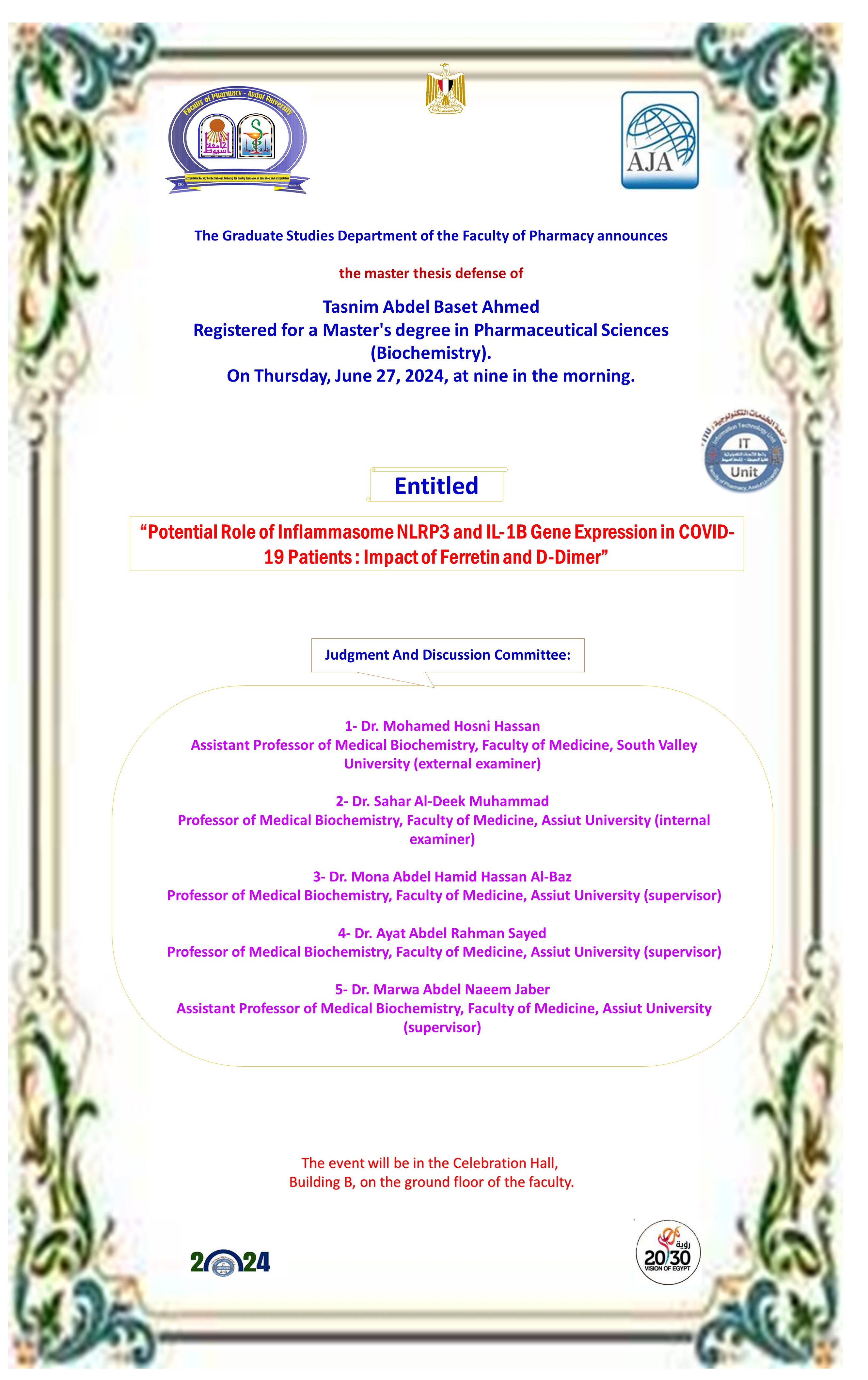
The thesis defense of the pharmacist Tasneem Abdel Basit Ahmed - registered for a master’s degree in pharmaceutical sciences (biochemistry) on Thursday, June 27, 2024
Meeting of the Executive Committee (Clinical Pharmacy Program) on Wednesday, July 10, 2024

God willing, the Executive Committee meeting will be held on This is Wednesday, July 10, 2024, at ten o’clock (AM).
The Faculty Council Hall - the fifth floor (administrative building)
Acting Dean of the Faculty
(Prof. Gihan Nabil Hassan Fetih)
Development of Highly Sensitive Fluorescent Sensors for Separation-Free Detection and Quantitation Systems of Pepsin Enzyme Applying a Structure-Guided Approach
Two fluorescent molecularly imprinted polymers (MIPs) were developed for pepsin enzyme utilising fluorescein and rhodamine b. The main difference between both dyes is the presence of two (diethylamino) groups in the structure of rhodamine b. Consequently, we wanted to investigate the effect of these functional groups on the selectivity and sensitivity of the resulting MIPs. Therefore, two silica-based MIPs for pepsin enzyme were developed using 3-aminopropyltriethoxysilane as a functional monomer and tetraethyl orthosilicate as a crosslinker to achieve a one-pot synthesis. Results of our study revealed that rhodamine b dyed MIPs (RMIPs) showed stronger binding, indicated by a higher binding capacity value of 256 mg g−1 compared to 217 mg g−1 for fluorescein dyed MIPs (FMIPs). Moreover, RMIPs showed superior sensitivity in the detection and quantitation of pepsin with a linear range from 0.28 to 42.85 µmol L−1 and a limit of detection (LOD) as low as 0.11 µmol L−1. In contrast, FMIPs covered a narrower range from 0.71 to 35.71 µmol L−1, and the LOD value reached 0.34 µmol L−1, which is three times less sensitive than RMIPs. Finally, the developed FMIPs and RMIPs were applied to a separation-free quantification system for pepsin in saliva samples without interference from any cross-reactors.
Enhanced dual fluorescence quenching of red and blue emission carbon dots by copper dimethyldithiocarbamate for selective ratiometric detection of ziram in foodstuff and water samples
The monitoring of ziram levels is of vital importance due to its widespread application in agriculture and the possible risks it poses to human health and the ecosystem. This work proposes an innovative approach for the highly sensitive and selective sensing of ziram, a widely used dithiocarbamate fungicide, through the formation of copper dimethyldithiocarbamate Cu)DDC)2 assisted dual quenching of red and blue emission carbon dots (R/NCDs and B/NSCDs). When ziram is added to a system containing copper-bound B/NSCDs and R/NCDs, the displacement of ziram zinc ions by copper ions leads to the formation of a yellow-colored Cu)DDC)2 complex. This complex induces significant quenching of the fluorescence emissions from both types of carbon dots, consequently enhancing the sensitivity of the detection method. Comprehensive characterization of the R/NCDs and B/NSCDs was conducted using various spectroscopic and morphological techniques to elucidate their structural and optical properties. The quenching mechanism was confirmed through different spectroscopic techniques, including fluorescence lifetime measurements, UV–vis spectroscopy, and Stern-Volmer analysis. The proposed method demonstrated high sensitivity across a wide concentration ranging from 20 to 1000 ng mL−1, making it suitable for trace-level detection of ziram. Moreover, the method exhibited excellent selectivity and recovery when applied to real samples, such as apples, grains, and water matrices. The combination of high sensitivity, selectivity, and applicability to diverse sample matrices makes this carbon dot-based dual quenching method a promising approach for fast and accurate detection of ziram in various environmental and agricultural applications.
Exploiting the pH-responsive behavior of zinc-dithizone complex for fluorometric urea sensing utilizing red-emission carbon dots
This study develops a novel fluorometric method for the sensitive and selective determination of urea, based on unique system comprising nitrogen doped red-emissive carbon dots (NRECDs), zinc-dithizone complex, and the urease enzyme. The underlying principle of this method relies on the pH increase resulting from the enzymatic breakdown of urea by urease. Initially, the fluorescence of the NRECDs is quenched by the red-colored zinc-dithizone complex. However, upon the addition of urea, the subsequent release of ammonia and the consequent rise in pH lead to the dissociation of the zinc-dithizone complex, causing a color change from red to yellow. This spectral shift eliminates the quenching effect, resulting in the restoration of the CDs’ fluorescence. The prepared NRECDs were comprehensively characterized using various spectroscopic techniques, including fluorometry, X-ray diffraction (XRD), X-ray photoelectron spectroscopy (XPS), UV–visible spectroscopy, and transmission electron microscopy (TEM) imaging. The proposed fluorometric method exhibits excellent sensitivity (Limit of detection = 0.0012 mM) and linearity (R2 = 0.9951) in the determination of urea. Notably, this approach addresses the selectivity limitations of previous pH-sensitive CDs-based methods, which relied solely on the intrinsic response of CDs, lacking specificity in either quenching or fluorescence enhancement. Furthermore, the developed method demonstrates remarkable selectivity, as evidenced by negligible interference from various potentially interfering substances, ensuring reliable and accurate urea quantification. When applied to human serum samples, the method showcased excellent recovery with low relative standard deviations, highlighting its practical applicability in biomedical and clinical applications.


