 Do you have any questions?
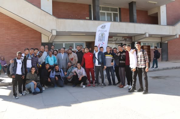

congratulations
Prof. Dr. Osama Al-Ashir for his effective participation in issuing child nutrition protocols for the Ministry of Health and Population in the Arab Republic of Egypt.

congratulations
Prof. Dr. Osama Al-Ashir for his effective participation in issuing child nutrition protocols for the Ministry of Health and Population in the Arab Republic of Egypt.
Corrective spine s urg eries for scoliosis and/or kyphosis are a major challenge. This study was carried out to test whether preoperative intravenous administration of amantadine sulfate could affect the intraoperative hemodynamics in patients undergoing major spine corrective surgeries
Introduction: Trichomoniasis remains one of the most significant sexually
transmitted disease (STDs) for public health. The disease is caused by parasitic
protozoa, Trichomonas vaginalis (T. vaginalis), which is often underestimated in
tropical medicine. Despite its public health importance, the epidemiology and
molecular characteristics of trichomoniasis in Egypt remains poorly understood,
particularly in the southern part of the country (Upper Egypt). This study targeted
exploring the genetic variability of T. vaginalis infections in Egyptian women
living in Upper Egypt using restriction fragment length polymorphism (RFLP).
Patient and techniques: This cross-sectional study included 150 female patients,
who visited the gynaecology and obstetrics outpatient clinics at Sohag General
Hospital between 2019 and 2022, exhibiting symptoms of trichomoniasis.
Vaginal washout samples were collected from each patient and analyzed using
three diagnostic techniques: direct wet mount microscopy, culture on TYM
Diamond’s medium, and PCR amplification and Polymerase Chain Reaction-
Restriction Fragment Length Polymorphism (PCR-RFLP) targeting the actin
gene, which was applied to all 16 samples that tested positive in culture. The
PCR-RFLP results were then visualized through agarose gels electrophoresis to
detect DNA fragments.
Results: Out of 150 vaginal washout samples, 12 cases (8%) tested positive for
T. vaginalis trophozoites via direct wet mount microscopy, while 16 samples
(10.6%) were positive in culture. Additionally, PCR-RFLP analysis of the 16
culture-positive samples revealed that 13 samples were confirmed positive
using this molecular method. The amplified products were digested with the
restriction enzyme Hind II, yielding three DNA fragments of 60, 213, and 827 bp,
which were then detected by agarose gel electrophoresis. Digestion with RsaIproduced five fragments measuring 87, 103/106, 236, and 568 bp, while MseI digestion resulted in three distinct fragments of 204, 315, and 581 bp.
Conclusion: This study provides robust baseline data on the prevalence and microscopic characteristics of T. vaginalis in Upper Egypt, while also presenting, for the first time, molecular detection and genotyping and revealed that genotype E is the only prevalent genotype in the region.
Aim: This research is a descriptive epidemiological study aimed to investigate the patient reported outcomes and expression of chronic pelvic pain of different etiologies and difference between males and females regarding the impact of pain on different life aspects including social and economic aspects.
Patient & method: This study was a descriptive analysis including patients diagnosed with chronic pelvic pain who visited pain management clinic at Assiut University hospital between 2018 and 2023. Five hundred patients diagnosed with CPP were allocated into two groups: 140 males and 360 females. All cases were monitored to assess the visual analog scale (VAS), PESQ, and health VAS scores. During the follow-up, the patients' efficacy evaluation and the administration of analgesics were documented.
Results: Regarding the type of pelvic pain in females, it was gynecological in 46.11% of patients, urological in 24.44% of patients; VAS was not significantly different between both patients. PSEQ was markedly elevated in male cases in comparison to female cases, with a P value of less than
Russell–Silver syndrome (RSS) is an uncommon but well-known imprinting condition primarily characterized by postnatal development failure and idiopathic intrauterine growth retardation (IUGR) and an inverted triangular face and a prominent forehead with relative macrocephaly that distinguish it from idiopathic IUGR and other causes of postnatal growth failure. Few case reports of RSS with cleft palate have been published and those who have perioperative issue such difficult intubation owing to trismus and difficulty to use a mouthpiece due to mandibular development failure.
Case presentation
Female child with RSS was subjected to cataract surgery performed under general anesthesia. Despite limited mouth opening and short thyro-mental distance, the intubation was relatively easy. The patient was extubated and moved to the postoperative care area. Postoperative interval passed uneventful …