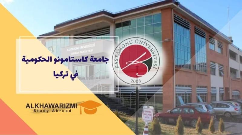
Merry Christmas greetings

تتقدم 

السيدة ا.د/ ايناس احمد عبد الحافظ

والسادة الوكلاء والسادة أعضاء هيئة التدريس والهيئة المعاونة والجهاز الإداري 

بخالص التهاني القلبية الى الاخوة الاقباط

أعضاء هيئة تدريس -هيئة معاونة - طلاب - جهاز ادارى

بعيد الميلاد المجيد

ونسأل الله تبارك وتعالي ان ينعم على مصرنا الحبيبة بنعمة الأمن والأمان
 

An Overview of Camel Brucellosis and Its Impact on Human Health in Endemic Regions

Research Abstract

Background: Camel brucellosis is a zoonotic disease caused by the genus Brucella and has significant economic and public health effects worldwide, particularly in areas where camels play a significant role in the pastoral economy. This review describes the epidemiological aspects of camel brucellosis and its impact on human health.

Methods: A comprehensive literature search was performed using PubMed, Web of Science, Scopus, and Google Scholar databases to locate studies on camel brucellosis for recognizing its epidemiological aspects and impact on human health. The search included 274 documents.

Results: The disease results in reduced milk output, reproductive failures, arthritis, and financial losses for farmers. The seroprevalence of camel brucellosis across the camel-rearing countries is alarming and ranges between 0.4 and 37.5%. Sudan and the Kingdom of Saudi Arabia are the most affected countries with camel brucellosis. Three species were isolated from diseased camels, including B. abortusB. melitensis, and B. suis. The potential risk factors of camel brucellosis include age, body condition, gender, physiological status and breed of the camel, herd size, contact with cattle and small ruminants, management, history of abortion, locality, and season. Accurate diagnosis of camel brucellosis is essential for effective management of the disease. Serodiagnosis is still one of the most used techniques for identifying the affected animals.

Conclusions: A one-health strategy is required from the governments, animal health professionals, and other stakeholders to combat the disease. This strategy includes enhancing veterinary care, increasing public and herders awareness toward the zoonotic importance of the disease as well as value of biosecurity procedures, and putting in place efficient immunization programs aimed at high-risk groups. Due to the socioeconomic impact of camel brucellosis, it is crucial to understand its epidemiology and potential risk factors to implement effective control strategies.

 Keywords:

Brucella spp.; complement fixation test; dromedary; enzyme-linked immunosorbent assay; Rose Bengal plate test; seroprevalence

Research Authors
Ahmed Abdel-Rady, Hanin Ashraf Abu-Seida and Ashraf M. Abu-Seida
Research Date
Research Department
Research File
Research Journal
Vector-Borne and Zoonotic Diseases
Research Member
Research Publisher
Scopus
Research Rank
Veterinary Microbiology
Research Website
https://doi.org/10.1177/15303667251380273
Research Year
2026

Morphomolecular Characterization of Rhadinorhynchus niloticus (Acanthocephala: Rhadinorhynchidae) from Nile Perch (Lates niloticus, Perciformes: Latidae)

Research Abstract

Adults of Rhadinorhynchus niloticus, a member of the Rhadinorhynchidae family, were isolated from the intestines of wild Nile perch (Lates niloticus (Linnaeus, 1758); Perciformes: Latidae) caught from the River Nile and its tributaries in Assiut City, Egypt. The parasite was found freely in the intestinal lumen with a prevalence of 10.71%, and the burden varied from one to five parasites per fish. The mean intensity and abundance were 2.16 ± 0.47 (95% CI: 1.33 to 3.17) and 0.23 ± 0.08 (95% CI: 0.11 to 0.43), respectively. The parasite was described using light and scanning electron microscopy. Molecular species identification as well as phylogenetic relationship analysis of the isolated parasite were achieved by sequencing and comparisons of the mitochondrial cytochrome oxidase C subunit I (COI) and nuclear 18S rRNA genes. The sequences were deposited in GenBank under the accession numbers PP859185 and MZ727194. Furthermore, phylogenetic analysis demonstrated that the parasites emerged from a separate branch belonging to the Rhadinorhynchidae family, which was clearly distinguished from other genospecies.

Research Authors
Marco Albano and Hasnaa Thabit Ebtsam Sayed Hassan Abdallah
Research Date
Research Journal
Fishes
Research Year
2025

Impact of Amirthalingamia macracantha Larvae on Nile Tilapia (Oreochromis niloticus): A Morpho-Histopathological Perspective

Research Abstract

Amirthalingamia macracantha (Joyeux and Baer, 1935) larvae, a member of the Gryporhynchidae family (Cestoda: Cyclophyllidea), are commonly found in a variety of African fish species, including Nile tilapia (Oreochromis niloticus, Linnaeus, 1758). Nonetheless, little is known regarding their existence in native Egyptian fish species. Two hundred O. niloticus were examined in the current investigation, using standard parasitological techniques to detect the presence of these larvae in different fish organs. According to this study’s findings, A. macracantha larvae were found in 35% of the microscopically examined samples as red nodules, attaching to the serosa of both the anterior and posterior sections of the intestine and stomach, respectively. The mean abundance of the infection was 3.5 ± 0.05 (CI 95% 3.4–3.5), and its mean intensity was 10 ± 1.7 (CI 95% 8.2–11.7). The existence of two rows of twenty rostellar hooks of varying lengths is the main characteristic of the parasite. The upper row consisted of four large hooks and six smaller ones, while the lower row contained smaller hooks of consistent length. According to histopathological analysis, the larvae with a hook-and-spiny rostellum were affixed to the stomach serosa of O. niloticus. Additionally, there was a notable presence of degranulated mast cells and lymphocytes of varying sizes within the stomach’s lamina propria, submucosa, and blood vessels, as well as a loss of gastric epithelium, fibrosis, and enlargement of submucosal lymph vessels. This study offers a succinct description of this larval stage, representing the first reported occurrence of its presence in Egyptian O. niloticus.

Research Authors
Sultan Mohammed Aresh Dalal Alardan and Yosra M. I. El Sherry Ebtsam Sayed Hassan Abdallah, Mahmoud Mostafa Mahmoud, Hanan Hassan Abdel-Hafeez, Marco Albano, Gioele Capillo, Asmaa Mohamed Metwally
Research Date
Research Journal
Animals
Research Year
2025

Molecular and epidemiological aspects related to bovine papular stomatitis in large ruminants in Assiut governorate, Egypt

Research Abstract

Bovine papular stomatitis (BPS) is a widespread epitheliotropic viral disease affects ruminants of all ages and considered a serious risk to public health. In Assiut governorate, there is little information on BPS despite their financial losses. Thus, the goal of this study was to confirm diagnosis on a molecular base, describe the clinical findings of the disease, and study the relationship between some factors that may play a role in the spread of the disease. The present investigation was conducted on 39 cattle and 11 buffaloes that belonged to different villages in Assiut governorate, Egypt. Whole blood and oral lesions swabs samples were taken for laboratory testing. Semi-nested polymerase chain reaction (semi-nested PCR) had been used for detection of BPS virus (BPSV). The viral DNA was detected in 32 cattle and 8 buffaloes. The result of clinical examination indicated that the clinical signs of BPS were oral (ulcers in gum, palate, papillae & tongue), commissure, muzzle, and nostrils lesions with one or more of other clinical signs such as fever, diarrhea, enlarged superficial lymph nodes, corneal opacity, respiratory distress and skin lesions in studied animals. Studying the effect of some factors on the spread of the disease revealed that the prevalence was significantly higher in middle areas of the governorate than in other areas. To lower the prevalence of BPSV, it is recommended to avoid eating hard hay and reeds that cause small abrasion in oral cavity and aid increasing frequency of BPSV infection.

Research Authors
Fatma E. Mahran1 , Khaled A.S. El-Khabaz1,2, Osama A. Ali1 , Zainab M.A. Youssef1
Research Date
Research Department
Research File
BPS 2026.pdf (451.39 KB)
Research Journal
Journal of Advanced Veterinary Research
Research Pages
209-212
Research Publisher
Zainab Mohammed Ahmed Mohammed
Research Vol
16
Research Year
2026

Molecular detection and macroelements changes in cattle infected with bovine ephemeral fever virus in Assiut Governorate, Egypt

Research Abstract

Bovine ephemeral fever (BEF) is a dangerous febrile viral disease that affects cattle and causes large financial losses. The aims of this study were to study clinical findings of BEF virus (BEFV) infection, identify certain risk factors and evaluate infected cattle’s mineral changes. One hundred and seven cattle were used in this investigation. Whole blood and serum samples were collected for laboratory analysis. Reverse transcriptase polymerase chain reaction (RT-PCR) had been employed for BEFV diagnosis. The observed clinical findings of BEFV infection in cattle were fever, respiratory manifestation (serous nasal discharge & cough), lacrimation, lameness, enlarged of superficial lymph nodes, drop of milk production, ruminal stasis, and recumbency. Of the studied diseased cattle, 43 (43%) of 100 whole blood samples showed molecularly positive results for G1 gene. The infection rate of BEFV had no significant variation by locality, age, sex, breed and season in the studied diseased cattle. Serum calcium level of confirmed BEFV positive samples was significantly lower (p<0.001) than serum calcium level in clinically healthy ones. Serum phosphorus and magnesium levels in BEFV infected cattle did not differ significantly (P<0.05) from that of clinically healthy ones. The importance of establishing efficient prevention and control strategies in Egypt must be emphasized in order to reduce the prevalence of BEFV infection in cattle.

Research Authors
Fatma S. Mahmoud1 , Heba A. Nasr2 , Mohammed G.M. Metwally3 , Amr S. Mahmoud4 , Fatma A. Shaaban5 , Zainab M.A. Youssef
Research Date
Research Department
Research File
BEF 2026.pdf (500.38 KB)
Research Journal
Journal of Advanced Veterinary Research
Research Pages
263-267
Research Publisher
Zainab Mohammed Ahmed Mohammed
Research Vol
16
Research Year
2026

Nebivolol rescued the liver and kidney from the coadministration of rivaroxaban and cisplatin by targeting inflammation, oxidative stress, and apoptosis in rats

Research Abstract

Cisplatin is among the most frequently utilized drugs for addressing malignant tumors, yet it can lead to organ harm, especially hepatotoxicity and nephrotoxicity. Furthermore, the anticoagulant rivaroxaban could poten­ tially cause injury to the liver and kidneys. This research aimed to examine the protective benefits of nebivolol, known for its pleiotropic and tissue-protective characteristics, against the harmful effects of rivaroxaban and cisplatin on the liver and kidneys. Male rats received cisplatin and/or rivaroxaban, and we evaluated hepato­ toxicity and nephrotoxicity by measuring serum concentrations of AST, ALT, LDH, albumin, bilirubin, creatinine, and blood urea. We also measured MDA, GSH, GPx, NO, TNF-α, and IL-6 in kidney and liver homogenates. Histopathological analysis was performed on liver and kidney tissue sections, and immunohistochemical detection of caspase 3 in liver tissue and NF-κB in kidney tissue was conducted. Our findings demonstrated that nebivolol supported the preservation of the liver and kidney structure and function by reducing the biochemical and pathological alterations caused by cisplatin and rivaroxaban. Nebivolol decreased the elevations in MDA, TNF-α, and IL-6 levels while maintaining GSH, GPx, and NO levels in liver and kidney tissues. Moreover, nebivolol lowered the levels of caspase-3 in the liver and NF-κB in the kidneys. In conclusion, our study indicates that nebivolol protects the liver and kidneys from the detrimental effects of cisplatin and rivaroxaban. 
 

Research Authors
Ahmed M. Abd-Eldayem, Marwa F. Ali, Esraa A. Ahmed
Research Date
Research Department
Research Journal
International Immunopharmacology
Research Member
Research Publisher
Elsevier
Research Vol
153
Research Website
https://www.sciencedirect.com/science/article/abs/pii/S156757692500476X
Research Year
2025

Valproate attenuates neuroinflammation and glial activation in a rat model of fibromyalgia: Evidence for antioxidant and nociceptive modulation

Research Abstract

Fibromyalgia syndrome (FMS) is a chronic, multidimensional musculoskeletal condition distinguished by severe nociceptive dysfunction, persistent fatigue, sleep disruptions, cognitive deficits, and emotional instability. Although valproic acid (VPA) has been used to treat epilepsy and bipolar disorder, its efficacy in altering neuropathic pain pathways remains unclear. In this investigation, we assessed the neuromodulatory characteristics of VPA (300 mg/kg, intraperitoneally) in an established FMS rat model, with a focus on neuroinflammation, oxidative stress, and glial activation. Behavioral evaluations for thermal hyperalgesia (paw withdrawal latency, PWL) and mechanical allodynia (paw withdrawal threshold, PWT) were performed at baseline (day 0), after induction (day 5), and at various intervals following VPA administration. Neurochemical evaluations demonstrated that VPA markedly diminished FMS-induced elevations in malondialdehyde (MDA), nitric oxide (NO), and pro-inflammatory cytokines such as tumour necrosis factor-α (TNF-α), interleukin (IL)-1β, and interleukin (IL)-6, while restoring antioxidant defences, such as glutathione (GSH) and superoxide dismutase (SOD). Histopathological examination demonstrated reduced neuronal degeneration and decreased immunoreactivity of glial fibrillary acidic protein (GFAP). These findings indicate that VPA reduces FMS-related pain and neuroinflammatory characteristics through antioxidative and glial-modulating mechanisms, indicating its potential for therapeutic repurposing in neuropathic pain syndromes 
 

Research Authors
Esraa A. Ahmed , Rasha B. Abd-ellatief , Marwa F. Ali , Ahmed M. Abd-Eldayem
Research Date
Research Department
Research Journal
Brain Research
Research Member
Research Pages
1-12
Research Publisher
Elsevier
Research Rank
Q2
Research Vol
1869
Research Website
https://www.sciencedirect.com/science/article/abs/pii/S0006899325005967
Research Year
2025

Surface Roughness and Cleanability: Evaluating the Impact of Magnetic Abrasive Finishing on Dairy Equipment

Research Abstract

Stainless steel tubes and pipes are vital in dairy processing but require frequent cleaning, leading to notable energy consumption and environmental impact. This study delves into the cleanability of wet milk deposits at temperatures of 40°C and 60°C on an exceptionally smooth internal surface achieved through magnetic abrasive finishing (MAF) with a surface roughness of 0.01 μm Ra. We compare this surface with non-MAF tubes having surface roughness values of 0.37 and 3.7 μm Ra. To assess cleaning effectiveness, the study measured milk and protein residue removal after deposition and cleaning processes, employing a cleaning solution flow pattern at Reynolds numbers (Re) of 659–1318. Results indicate that smoother surfaces, particularly those with roughness values of 0.01 and 0.37 μm Ra, significantly enhance cleanability at 40°C. This leads to reductions (p < 0.05) of 34.5% and 22.6% for milk deposits and 27.9% and 22.7% for protein deposits, respectively, compared to tube surfaces with a roughness level of 3.7 μm Ra. These findings underscore the potential of highly smooth surfaces to improve cleanability below protein denaturation temperatures. Furthermore, the MAF tube with a roughness of 0.01 μm Ra exhibited nonsignificant reductions of 15.4% and 6.7% compared to the 0.37 μm Ra surface. The smoothing effect on the cleanability of milk and protein deposits was enhanced compared with the higher temperature condition. By addressing the challenges of routine cleaning, the study highlights MAF as a technology that optimizes surface quality in dairy processing equipment, addressing environmental and energy-related concerns.

Research Authors
Ikko Ihara, Hiroki Tokuda, John K. Schueller, Israa M. A. Mohamed, Yushi Sakamoto, Kiyohiko Toyoda, Kazutaka Umetsu, Hitomi Yamaguchi
Research Date
Research Department
Research Journal
Journal of Food Process Engineering i
Research Member
Research Pages
e70106
Research Publisher
Wiely
Research Rank
4
Research Vol
48
Research Website
https://onlinelibrary.wiley.com/doi/abs/10.1111/jfpe.70106
Research Year
2025

Cytomorphology, osmotic fragility, glucose-6-phosphate dehydrogenase and oxidant/antioxidant status in postparturient haemoglobinuria in dairy cattle and buffaloes

Research Authors
Ahmed Hassanin, M Karam, Nasreldin M Aref
Research Date
Research Department
Research Journal
Bulgarian Journal of Veterinary Medicine
Research Pages
55
Research Publisher
Faculty of Veterinary Medicine, Trakia University, Stara Zagora, Bulgaria
Research Vol
28
Research Website
https://bjvm.trakia-uni.bg/
Research Year
2025
Subscribe to