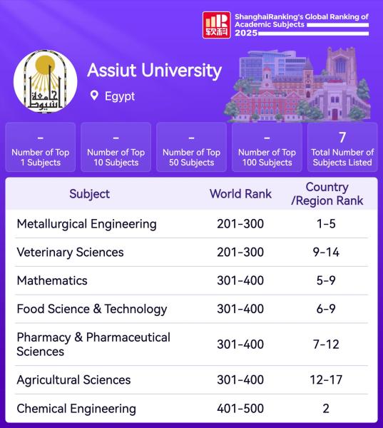
تصنيف شنغهاي
A commendable achievement added to the record of the Faculty of Veterinary Medicine - Assiut University, after its appearance in the Shanghai Ranking 2025
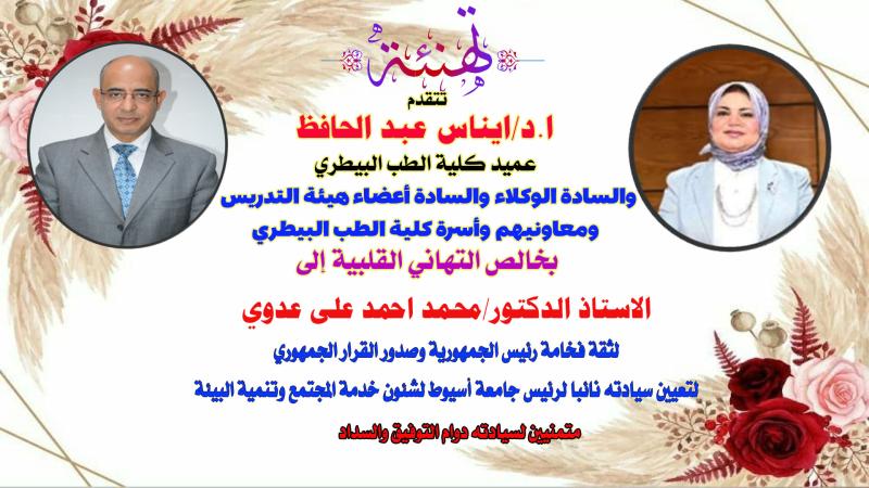
Congratulations from the Dean and faculty of the Faculty of Veterinary Medicine to Professor Mohamed Ahmed Ali Adawy on his appointment as Vice President of Assiut University for Community Service and Environmental Development.
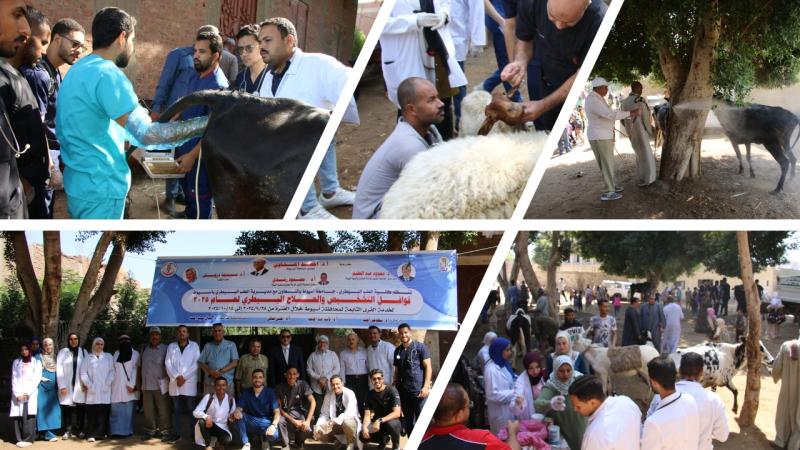
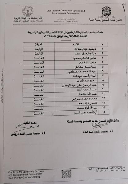
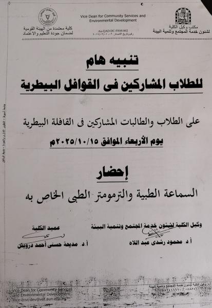
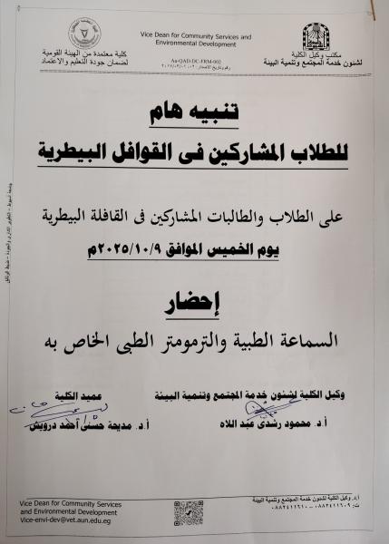
The Faculty of Veterinary Medicine at Assiut University is conducting a free veterinary caravan to the village of Bahij on Wednesday, October 15, 2025.
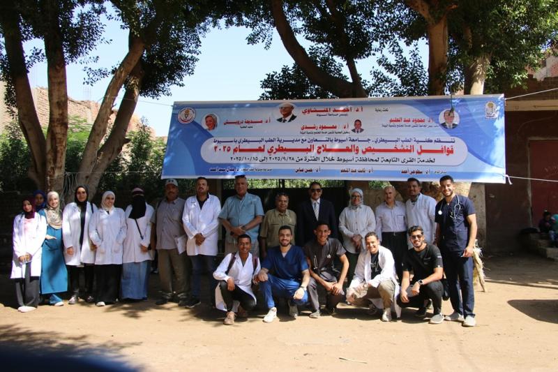
كلية الطب البيطري بجامعة أسيوط تنفذ قافلة بيطرية مجانية الى قرية بهيج يوم الأربعاء 15 أكتوبر 2025
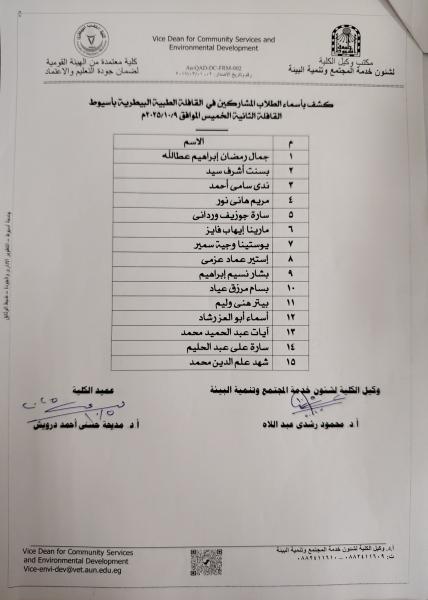
تحت رعاية الدكتور أحمد المنشاوي رئيس جامعة أسيوط والدكتور محمود عبد العليم نائب رئيس الجامعة لشئون خدمة المجتمع وتنمية البيئة والدكتورة مديحة درويش عميد الكلية والدكتور محمود رشدي وكيل الكلية لشئون خدمة المجتمع وتنمية البيئة، تم تنفيذ قافلة بيطرية مجانية يوم الأربعاء 15 أكتوبر 2025 الى قرية بهيج التابعة لمحافظة أسيوط، هذا وقد تم تنفيذ عدد من القوافل البيطرية المجانية إلى القرى التابعة لمحافظة أسيوط خلال شهري سبتمبر واكتوبر 2025، حيث تم تنفيذ قافلة بقرية شطب يوم 28 سبتمبر وقافلة الى قرية الزاوية يوم 9 أكتوبر، وتم تنفيذ هذه القوافل تحت رئاسة واشراف الدكتورة مديحة درويش عميد الكلية وتحت إشراف الدكتور محمود رشدي وكيل الكية لشئون خدمة المجتمع وتنمية البيئة والدكتور مؤمن عبد العظيم وكيل الكلية لشئون الطلاب وبمشاركة نخبة من أعضاء هيئة التدريس والهيئة المعاونة والطلاب والعاملين بالكلية.
في إطار الجهود المضنية والفعالة لإرساء مختلف أوجه المشاركة المجتمعية كأحد المحاور الأساسية للإستراتيجية الوطنية للتعليم العالي والبحث العلمي 2030 وفي ظل جهود النهوض بالثروة الحيوانية ونشر الثقافة البيطرية والوعي البيطري ومجابهة المسببات المرضية المختلفة للحيوان والدواجن والتي تشكل خطرا على صحة الإنسان والحيوان وفي سبيل السعي إلى تحقيق أهداف التنمية المستدامة، انطلقت فاعليات القافلة البيطرية التي نفذتها كلية الطب البيطري بجامعة أسيوط.
شارك في فاعليات القافلة البيطرية إلى قرية بهيج الدكتور محمود رشدي وكيل الكلية لشئون خدمة المجتمع وتنمية البيئة والدكتور مصطفى محمد احمد الأستاذ المتفرغ بقسم صحة الحيوان والدواجن والصحة البيئية والدكتور حسن عباس أستاذ ورئيس قسم التغذية والتغذية الإكلينيكية وعميد الكلية الأسبق والدكتور أحمد عبد الباقي الأستاذ المتفرغ بقسم الطب الشرعي والسموم البيطرية والدكتورة فاطمة محمود الأستاذ بقسم السلوكيات ورعاية الحيوان والدواجن والدكتور محمد حمدي الأستاذ بقسم الجراحة والتخدير والأشعة والدكتورة عبير عبد الوارث المدرس بقسم طب الحيوان، هذا بالإضافة الى عدد 5 من المدرسين المساعدين والمعيدين بالإقسام الإكلينيكية للكلية وعدد 20 طالب وطالبة بالفرق النهائية بالكلية، وتم في هذه القافلة فحص وعلاج عدد 1344 من الحيوانات المختلفة والطيور.
وتساهم مديرية الطب البيطري بأسيوط في التنسيق للقوافل البيطرية باختيار القرى الأكثر احتياجا وفي القيام بالأمور المتعلقة بالإعلان المسبق عن القافلة بالقرى مما يسهم في توافد عدد أكبر من الحالات المريضة وخدمة عدد أكبر من مربي الحيوانات.
وفي نهاية القافلة عبر مربي الحيوانات عن امتنانهم وشكرهم لجامعة أسيوط ولكلية الطب البيطري لتوفير الدعم الكامل لإقامة القوافل البيطرية والتي ترفع عن كاهلهم التكاليف الباهظة للتشخيص والعلاج، مما يساهم بكفاءة وفاعلية في تنمية الثروة الحيوانية وزيادة الإنتاج.