Professor Dr. Ahmed El-Menshawy, President of Assiut University, congratulates Professor Dr. Enas Abdel-Hafez on the issuance of the presidential decree appointing her as Dean of the Faculty of Veterinary Medicine.
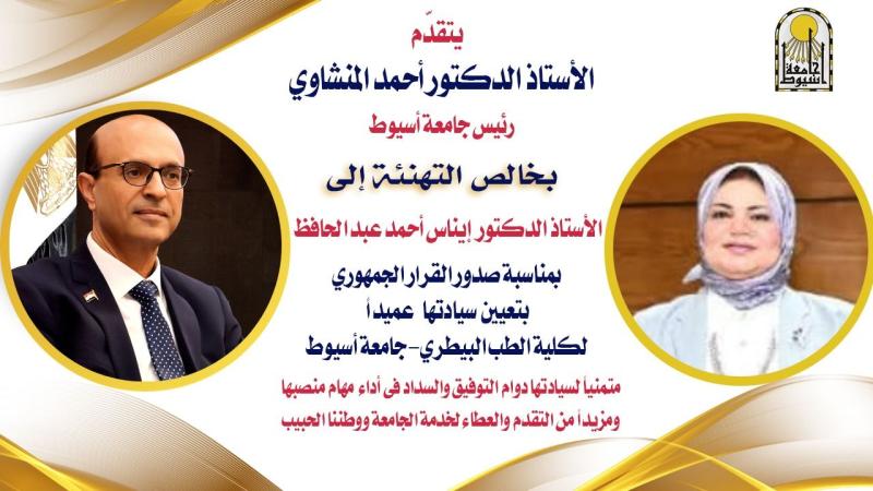
الدكتور المنشاوي يهنئ الدكتورة إيناس عبدالحافظ بمناسبة صدور القرار الجمهوري بتعيينها عميدةً لكلية الطب البيطري
قدّم الدكتور أحمد المنشاوي، رئيس جامعة أسيوط، خالص التهاني إلى الدكتورة إيناس أحمد عبدالحافظ بمناسبة صدور القرار الجمهوري بتعيينها عميدةً لكلية الطب البيطري، متمنيًا لها دوام التوفيق والسداد في مهام منصبها الجديد، ومواصلة مسيرة التطوير والارتقاء بالمنظومة التعليمية والبحثية داخل الكلية، وتعزيز دورها في خدمة المجتمع وتنمية البيئة.
وأعرب رئيس الجامعة عن خالص التقدير والعرفان لفخامة الرئيس عبد الفتاح السيسي، رئيس الجمهورية، وللأستاذ الدكتور أيمن عاشور، وزير التعليم العالي والبحث العلمي، تقديرًا لما يوليه فخامة الرئيس من اهتمام ورعاية لتطوير منظومة التعليم العالي والبحث العلمي في مصر، وما يبذله معالي الوزير من جهود حثيثة لدعم الجامعات المصرية، وتطوير بنيتها المؤسسية، وتعزيز تنافسيتها على المستويين الإقليمي والدولي.
وأكد الدكتور المنشاوي أن كلية الطب البيطري تُعد من الصروح العلمية المتميزة بجامعة أسيوط، إذ تضطلع بدور محوري في إعداد كوادر بيطرية مؤهلة علميًا وعمليًا، قادرة على خدمة المجتمع وتنمية الثروة الحيوانية، ودعم البحث العلمي والتطبيقي في مجالات الطب البيطري وسلامة الغذاء وصحة الحيوان والإنسان، بما يواكب احتياجات سوق العمل ويسهم في تحقيق رؤية الجامعة نحو التميز وخدمة المجتمع.
وأشاد رئيس الجامعة بما تتمتع به الدكتورة إيناس عبدالحافظ من كفاءة علمية، وخبرة أكاديمية وإدارية متميزة، تؤهلها لقيادة الكلية نحو مزيد من التطوير والريادة.
وأشار إلى أن جامعة أسيوط تؤمن بأهمية العمل بروح الفريق الواحد، وتحرص على تعزيز التواصل والتكامل بين مختلف كلياتها وقطاعاتها لتحقيق أهدافها الاستراتيجية في التميز الأكاديمي والبحثي وخدمة المجتمع.
والجدير بالذكر أن الدكتورة إيناس أحمد عبدالحافظ تخرّجت في كلية الطب البيطري بجامعة أسيوط عام 1994، وحصلت على درجة الماجستير في علم الأنسجة عام 1998، ثم نالت درجة الدكتوراه من الكلية نفسها عام 2004، وتدرجت في المناصب الأكاديمية حتى نالت درجة الأستاذية عام 2015. كما شغلت منصب وكيلة الكلية لشئون الدراسات العليا والبحوث قبل توليها العمادة، وأسهمت خلال مسيرتها العلمية في إثراء مجال تخصصها بعدد من الأبحاث والدراسات المتخصصة، إلى جانب إشرافها على العديد من رسائل الماجستير والدكتوراه.