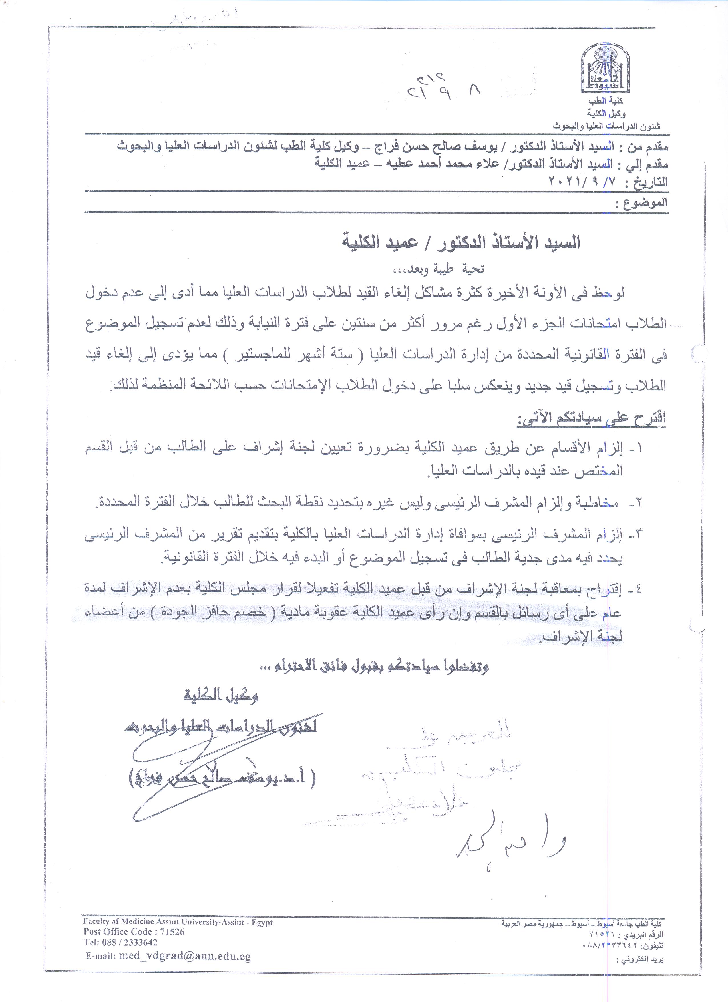
 Do you have any questions?
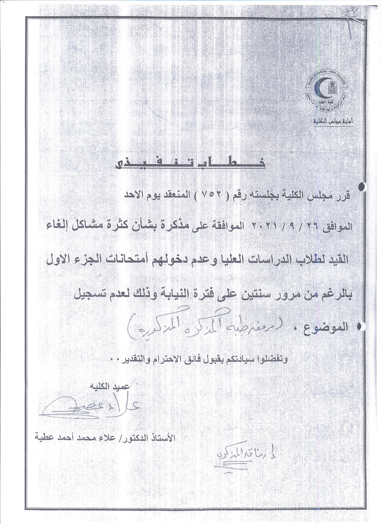
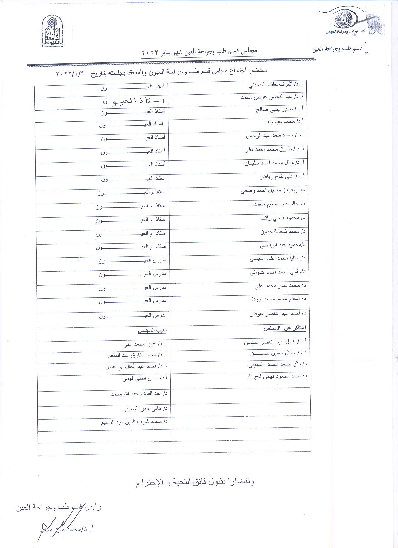
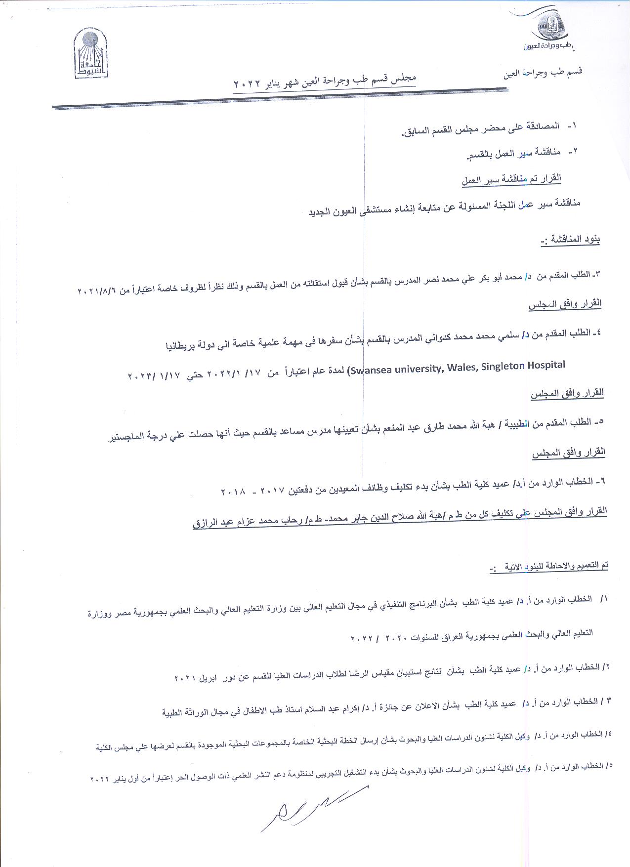
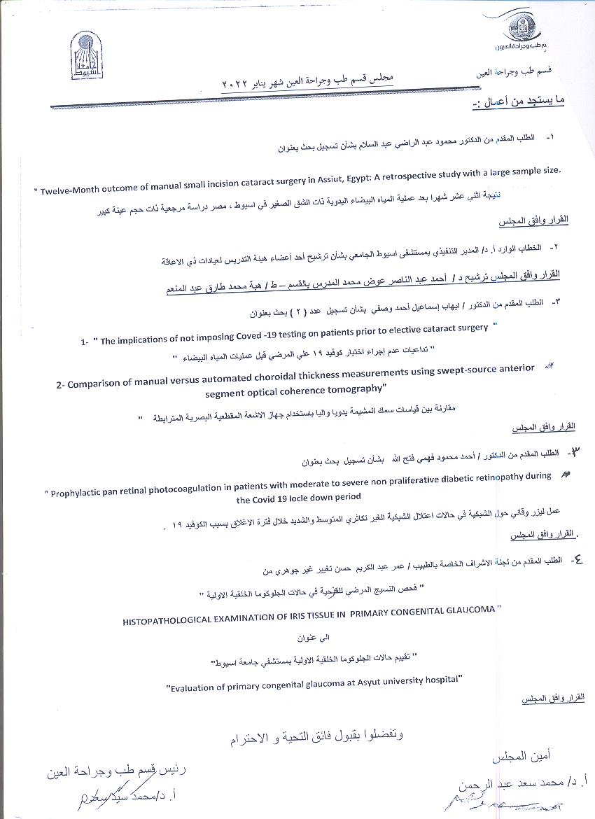
Department Board Minutes - Ophthalmology - January 2022
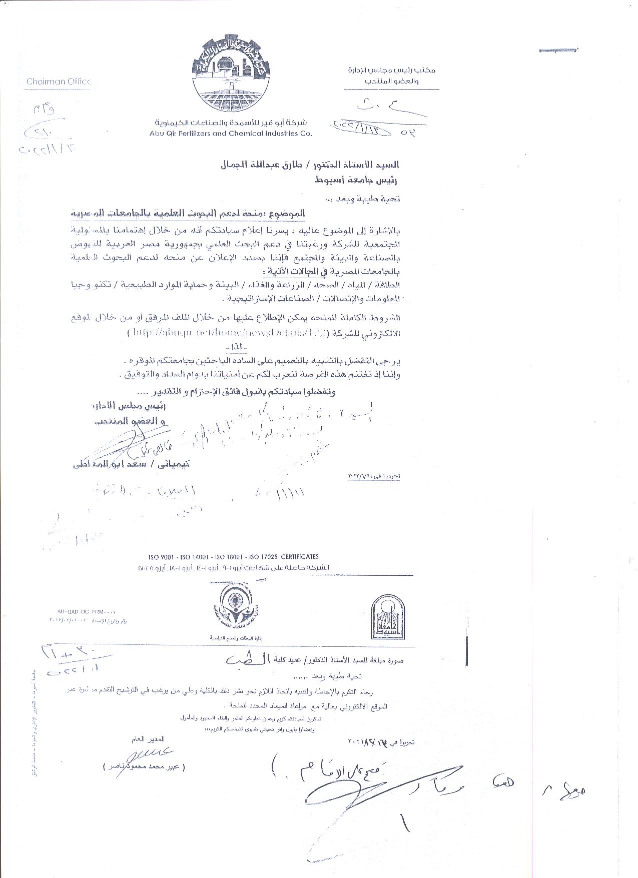
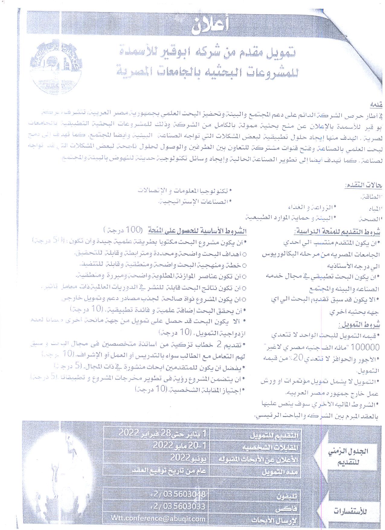
Funding provided by Abu Qir Fertilizer Company for research projects in Egyptian universities
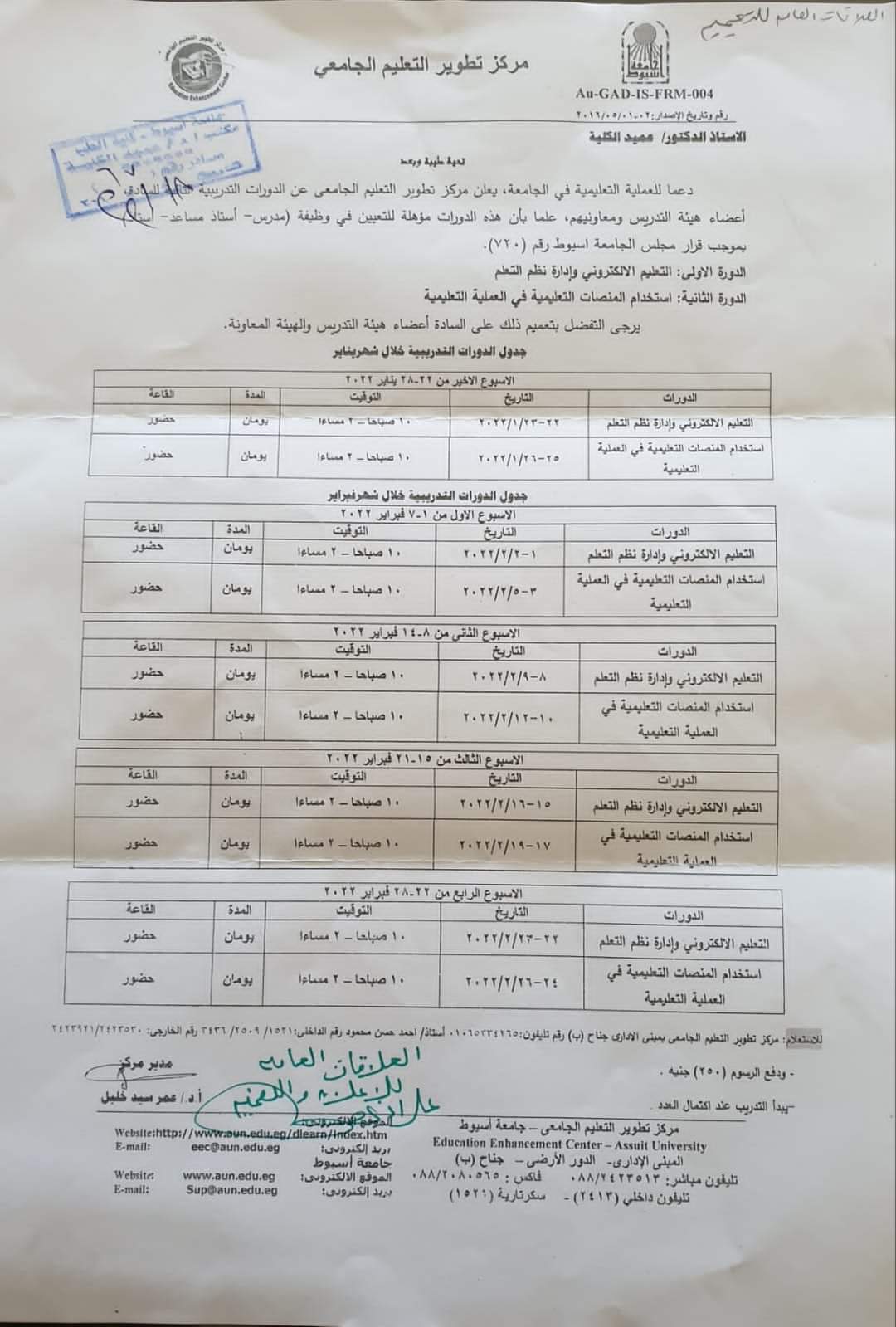
Schedule of training courses for the University Education Development Center for the month of January and February 2022
High Definition transcranial Direct Current (HD-tDCS) stimulation over the primary motor and insular cortex reduces capsaicin-induced pain and hyperalgesia
Research Abstract
There is an increasing interest in the use of non-invasive brain stimulation to modulate cortical activity for treating pain and psychiatric disorders. One such method is transcranial Direct Current Stimulation (tDCS) which has a reasonably high safety profile with few side effects and easy to administer. A number of studies reported that it can relieve neuropathic, musculo-skeletal and visceral pain as well as headaches and migraine. High Definition Transcranial Direct Current Stimulation (HD-tDCS) is a refinement of the tDCS method and aims to focus the area of stimulation to a specific brain region. We have tested this technique targeting a novel brain area in a human model of experimental pain. In a sham-controlled single-blinded trial we investigated the effects of HD-tDCS over two cortical targets: the primary motor cortex and the insula in a human model of capsaicin-induced pain.Thirty healthy volunteers (14 female, mean age 32.8 years) were divided into 3 groups and all had capsaicin cream (0.075%) applied for 30 minutes on a 9 cm2 area of skin on the volar surface of both arms. The areas of hyperalgesia after capsaicin application were measured by mapping the areas of primary and secondary hyperalgesia using a brush, a 19 g-Von-Frey Filament, and a calibrated 40 g-pinprick (Neurotips). Ten volunteers in each group then received 20 minutes of anodal HD-tDCS (2 mA) targeting the left primary motor cor-tex, left insular cortex or sham-placebo stimulation. Cortical targeting for stimulation was determined after running com-puter simulated brain conductivity models. Cutaneous areas of hyperalgesia were re-mapped after stimulation. The visual analogue pain scale was repeated throughout the experiment for assessing pain severity.We found a significant reduction in subjective pain scores (VAS) and areas of primary and secondary hyperalgesia after motor or insular cortex HD-tCDS compared to sham stimula-tion. The effects of insula stimulation appear to be bilateral while the reduction in cutaneous hyperalgesia was mainly contralateral to motor cortex HD-tCDS. We found that volun-teers in the stimulation groups also reported a significantly faster reduction in pain scores compared to the sham group. The volunteers failed to differentiate whether they had active or sham stimulation.In a human model of capsaicin-induced pain, anodal HD-tCDS of motor and insula cortex was shown to reduce pain and hyperalgesia compared to sham stimulation. This study shows that it is possible to stimulate the insular cortex using HD tCDS and the effects are bilateral as opposed to motor cortex stim-ulation where reduction in pain and hyperalgesia is predom-inantly contralateral.
Research Department
Research Journal
BJA
Research Member
Dexamethasone Versus Magnesium Sulfate As An Additives to Bupivacaine in Ultrasound Guided Supraclavicular Brachial Plexus Blockade
Research Abstract
September 2016 • Volume 123 • Number 3 Supplement
DEXAMETHASONE VERSUS MAGNESIUM SULFATE AS AN ADDITIVES TO BUPIVACAINE IN
ULTRASOUND GUIDED SUPRACLAVICULAR BRACHIAL PLEXUS BLOCKADE
R. A. Hamed1, N. M. Osman1, W. S. Hassan1,*, S. M. Omar1
1Anaethesia and Pain Management, Assuit University Hospital, Assuit, Egypt
Background & Objectives: comparing the effect of adding dexamethasone and magnesium
sulfate to bupivacaine in ultrasound guided supraclavicular brachial plexus blockade. The primary
endpoints were the onset of sensory and motor block and quality of analgesia and the
secondery end points were duration of analgesia and motor block.
Materials & Methods: Ninety patients scheduled for elective surgeries on the upper limb
under ultrasound guided supraclavicular brachial approach. patients were randomly allocated
using a standard randomization code, in a double blinded manner, into three groups; group one
(n=30) received 20 ml 0.5% plain bupivacaine, group two (n=30) received 18 ml bupivacaine
0.5% + 2 ml (8mg) dexamethasone, group three (n=30) received 18 ml bupivacaine 0.5% +
200 mg magnesium sulfate.
The following parameters were studied: Onset of sensory block, onset of Motor blockade,
haemodynamic and respiratory data, Analgesia time,and Duration of motor and sesory blocks
Results: There was no significant difference in the demographic data and surgical characteristics
in the three groups. The sensory block onset time in minutes was earlier in group 2
(dexamethasone) as compared to group 1 and 3; 8.20±2.09 versus 16±3.48 (P< 0.05) and
8.20±2.09 versus 12.70 ± 2.92(P< 0.05) respectively, also the sensory block onset was earlier
in group 3 (magnesium) than group 1 (control) 12.70 ± 2.92 versus 16±3.48 (P< 0.05).
Also onset of motor blockade in minutes was earlier in group 2 than in group 1 and 3; 1.50
± 2.09 versus 13.10 ± 3.34 (p< 0.05) and 12.75 ± 3.43 respectively, while the difference in
motor block onset was clinically insignificant between group 1 and 3(P> 0.05). As regard duration
of analgesia in minutes it was significantly longer in group 2 than group 1 and 3 1104.00
± 289.16 versus 313.50 ± 103.68 and 558.00 ± 48.08 respectively, also analgesia duration
was significantly prolonged in group 3 than group 1(P< 0.05). the motor power was significantly
prolonged for group 2 than group 1 and 3; 563.00 ± 69.29 versus 136.50 ± 28.34 and
214.50 ± 36.92 respectively, and also the motor power return was delayed in group 3 than
group 1(P< 0.05).
Conclusion: both of them proved to prolong the duration of block and the analgesia time, both
of them fasten the sensory block onset time, but dexamethasone is significantly more effective
in prolonging the analgesia duration and the block duration. dexamethasone shorten the
motor block onset time while magnesium does not enhance the motor block onset time.
Disclosure of Interest: None declared
DOI: 10.1213/01.ane.0000492692.50087.95
Research Date
Research Department
Research Journal
anesthesia and analgesia suplement
Research Member
Suppression effect of thyme and carvacrol nano-emulsions on Aspergillus fumigatus isolated from patients in the intensive care unit of Assiut
Research Abstract
Description
Background and Aim: Aspergillus fumigatus is a zoonotic fungus that causes several diseases in humans ranging from allergic reaction to fatal disseminated invasive infection, especially in immunocompromised patients. This study aimed to investigate the incidence of invasive A. fumigatus in patients admitted to the intensive care unit (ICU) of Assiut University Hospital, highlight the factors associated with their infection, and determine the antifungal effect of thyme nano-emulsion (TNE) and carvacrol nano-emulsion (CNE) on isolated A. fumigatus strains.
Materials and Methods: Mycological culture method and scanning electron microscopy (SEM) were used in the identification of A. fumigatus in 630 blood samples collected from 210 patients. TNE and CNE at five concentrations (1%, 2%, 4%, 6%, and 8%) and average sizes of 90.3 and 75.6 nm, respectively, were characterized by transmission electron microscopy. Their effect on A. fumigatus isolate growth was evaluated by the well-diffusion method and SEM, which was used for the detection of the degenerative effect of A. fumigatus ultrastructure.
Results: A. fumigatus was detected in 54 of 210 (25.7%) patients in the ICU. Advanced age and chronic diseases were considered important risk factors for invasive aspergillosis, especially in patients with more than 1 clinical disease. TNE and CNE showed an inhibitory effect on A. fumigatus isolates, which significantly increased with high concentrations. The respective values for TNE at concentrations of 6% and 8% were 6±0.41 mm and 15±0.67 mm. CNE completely inhibited A. fumigatus growth at concentrations of 4%, 6%, and 8%, while mean …
Research Date
Research Department
Research Journal
International Journal of One Health
Research Member
Fentanyl versus dexamethasone or both as adjuvants to bupivacaine in an ultrasound-guided paravertebral block in patients undergoing modified radical mastectomy
Research Abstract
Background
This study aimed to compare the effect of dexamethasone added to fentanyl and bupivacaine with the effect of either dexamethasone or fentanyl alone when combined with bupivacaine. in the thoracic paravertebral block (TPVB).
Methods
Sixty female patients (aged 18-60 years), scheduled for modified radical mastectomy were enrolled. Patients received preoperative unilateral paravertebral block using 0.3 ml/kg of 0.5% bupivacaine combined with 8 mg dexamethasone (Group I), 1 μg/kg fentanyl (Group II), or 8 mg dexamethasone+ 1 μg/kg fentanyl (Group III). The study drugs were diluted with normal saline 0.9% up to 25ml volume. The primary outcome was the time to first postoperative analgesics request, Secondary outcomes were total analgesic consumption, verbal rating pain scale (VRS) over the first 24 hours postoperatively, hemodynamic parameters, and adverse effects.
Results
The time to first analgesic request for intravenous (IV) nalbuphine was longer in group II (15.75±0.9 h, P< 0.001) than group I (10.45±1.1 h, P< 0.001), while no patients requested it in group III (P< 0.001). The total analgesic consumption of IV nalbuphine was lower in group II (8.6±3.5 mg, P= 0.04) than group I (11.3±2.1 mg), with a significant difference between group II and III (P< 0.001). From the 8th till the 24th hours postoperatively, patients in group III showed the significantly lowest median VAS scores, followed by patients in group II (P< 0.001) and lastly patients in group I. There were no significant adverse effects.
Conclusions
Research Department
Research Journal
Minerva Anesthesiol
Research Member
Elevated hippocampal mGlut2 receptors in rats with metabolic syndrome-induced-memory impairment, possible protection by vitamin D.
Research Abstract
Background
Metabolic syndrome patients are commonly prone to major health problems as cardiovascular diseases, diabetes mellitus, chronic kidney disease, cancer and neuropsychological complications including dementia.
Objectives
This research investigates mechanisms linking metabolic syndrome to cognitive impairment and possible impact of vitamin D supplementation.
Methods
Forty male Wistar rats were divided into 4 groups. Control, metabolic syndrome (20% fructose solution in drinking water for 12 weeks, vitamin D supplemented (500 IU/kg/day) and metabolic syndrome supplemented with vitamin D. Animals were assessed for spatial memory, hippocampal expression of SNAP 25, VAMP and mGlut2 receptor and hippocampus histological examination. Animals with metabolic syndrome showed prolonged acquisition and retention latencies in morris water maze, decreased hippocampal expression of …
Research Date
Research Department
Research Journal
Brain Research Bulletin
Research Member
Research Pages
108-117
Research Vol
180
Research Year
2022
Influence of chemically and biosynthesized silver nanoparticles on in vitroviability and infectivity of Trichinella spiralismuscle larvae
Research Abstract
ABSTRACT. Trichinellosis is a serious worldwide parasitic zoonosis. The available therapy for the treatment of
Trichinella spiralisis not satisfactory. Therefore, the recovery of effective treatment is required. This work aimed at
evaluating of the in vitro effect of silver nanoparticles (AgNPs) on muscle larvae of Trichinella. The present study
investigated the larvicidal properties of chemical and myrrh AgNPs on muscle larvae (ML) of T. spiralis. The used
AgNPs were chemically prepared using NaBH4
as reducing agentand biosynthesized using methanolic myrrh extract.
Characterization of synthesized AgNPs was monitored via UV-vis spectrophotometry, Fourier transform infrared
spectroscopy and transmission electron microscopy (TEM) studies. The ML incubated with AgNPs at concentrations
ranged from 1 μg/ml to 20 μg/ml. Chemical and biosynthesized AgNPs revealed marked larvicidal effect against ML of
Trichinella. Additionally, this in vitro study showed degenerative changes affecting the cuticle of AgNPs treated ML.
The effectiveness of AgNPs on the infectivity of TrichinellaML was also assessed. The results showed complete
inhibition of the infectivity of ML exposed to sublethal doses of chemical and myrrh prepared AgNPs when used to
infect animal models. This is the first report where myrrh synthesized AgNPs have been tested for their anthelminthic
activity against Trichinellain an in vitromodel.
Keywords: Trichinella spiralis, muscular larvae, silver nanoparticles, infectivity, viability
Research Date
Research Department
Research File
Research Journal
Annals of Parasitology
Research Member
Research Pages
591–602
Research Publisher
Polish Parasitological Society
Research Rank
international Impact factor 0.77
Research Vol
67(4)
Research Website
https://www.annals-parasitology.eu
Research Year
2022