 Do you have any questions?
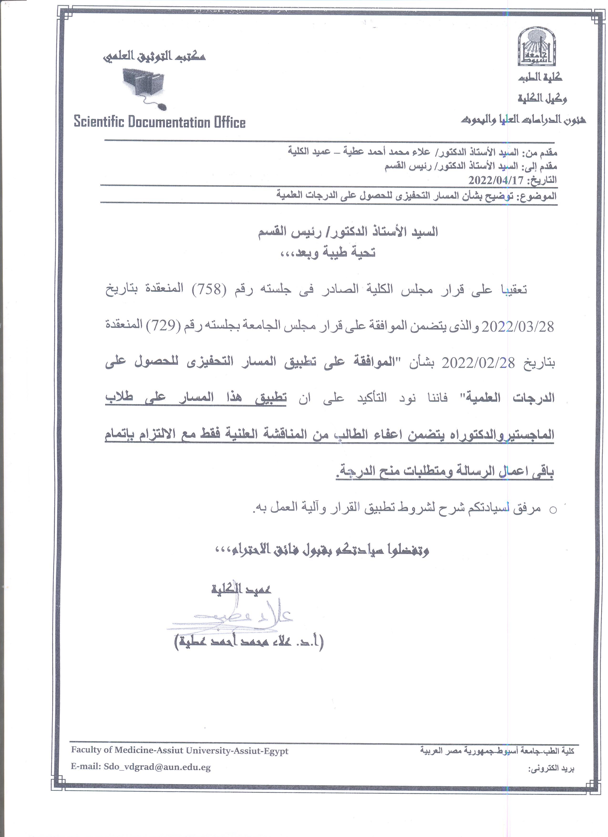
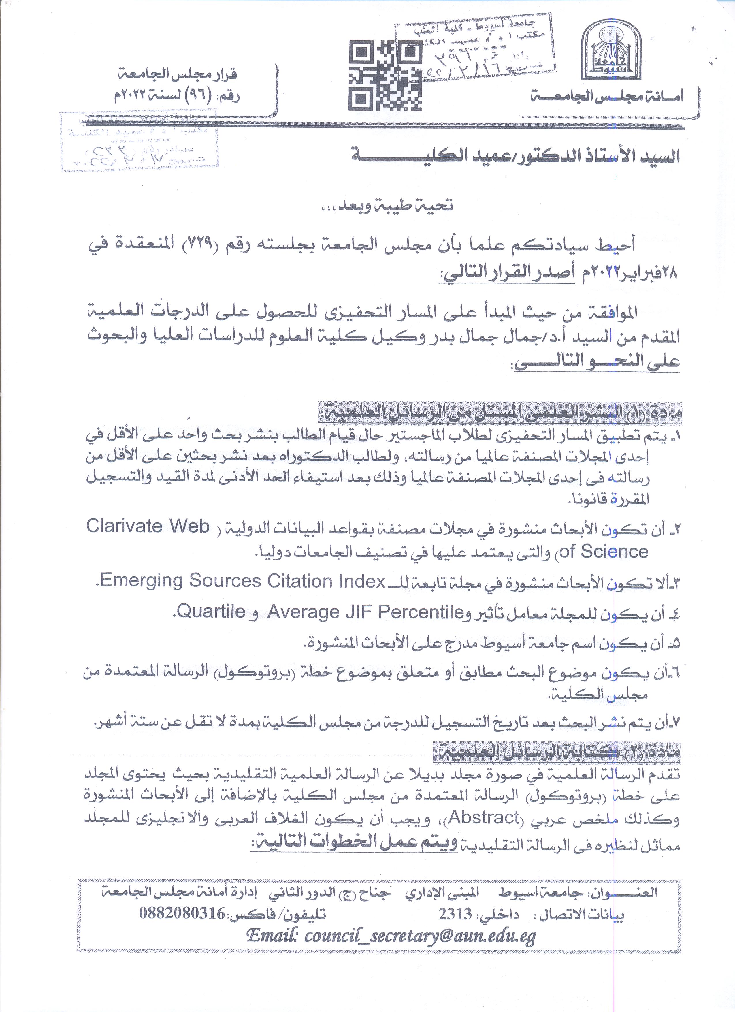
Clarification on the motivational path for obtaining scientific degrees
Comparison of high risk characteristics of non-culprit plaques in CCS vs ACS
Research Abstract
Most studies comparing lesion characteristic of non-culprit plaques of acute coronary syndrome (ACS) patients to plaques of stable angina patients studied only in hemodynamically insignificant nonculprit plaques (eg diameter stenosis <30 or 50%). With increase in plaque burden, plaque composition might change. Lipidic content measured by near-infrared spectroscopy (NIRS) and represented by lipid core burden index (LCBI) has demonstrated its correlation with future non-culprit major adverse cardiac events. New insights in plaque composition of hemodynamically significant non-culprit
lesions may serve to understand the potential of pharmacological therapy to prevent non-culprit major adverse cardiac events.
Research Date
Research Department
Research Journal
EuroPCR
Research Member
Research Year
2022
Employees Knowledge about Glaucoma at Assiut University Employees
Research Date
Research Department
Research Journal
Assiut Scientific Nursing Journal
Research Member
EPIDEMIOLOGICAL AND MORPHOLOGICAL STUDIES ON HYALOMMA SPECIES INFESTATING DROMEDARY CAMELS IN ASWAN GOVERNORATE, EGYPT
Research Abstract
Abstract The study evaluated the hard ticks’ prevalence parasitizing dromedary camels (Camelus dromedaries) in Aswan Governorate from July 2020 to August 2021. Of 1190 camels examined 1060 (89%) were infested. Factors were camel age, sex, infested site, and seasonal variations association with the tick distribution, as camels > 10 years were at higher risk rate of 97% (390/410). The infestation rate was 90% (990/1100) in males and 77.8% (70/90) in females. Exposure to infestation was higher in summer 93.7% (458/489) than other seasons. The face, udder, testes, hind limbs, and tail were the most infested site 97.5% (1160/1190) for each, followed by forelimbs was 96.6% (1150/1190), chest was 94% (1120/1190), and abdomen was 92.4% (1100/1190). Statistical analysis of the possible associated risk factors, camel’s age, gender, sampling season, and infested parts within animal body were all found to be significantly a ffected and related to hard tick distribution (P<0.05). Also, the identification and morphological characterization of the isolated hard ticks were performed using stereomicros
Research Date
Research Department
Research File
Research Journal
Journal of the Egyptian Society of Parasitology
Research Member
Research Pages
123 - 132
Research Publisher
the Egyptian Society of Parasitology,
Research Rank
INTERNATIONAL
Research Vol
52(1)
Research Website
https://jesp.journals.ekb.eg/
Research Year
2022
Adipose tissue-derived mesenchymal stem cells reduce endometriosis cellular proliferation through their anti-inflammatory effects
Research Abstract
Objective: Endometriosis is a chronic debilitating inflammatory condition characterized by the presence of endometrial tissues outside the uterine cavity. Pelvic soreness and infertility are the usual association. Due to the poor effectiveness of the hormone therapy and the high incidence of recurrence following surgical excision, there is no single effective option for management of endometriosis. Mesenchymal stem cells (MSCs) are multipotent stromal cells studied for their broad immunoregulatory and anti-inflammatory properties; however, their efficiency in endometriosis cases is still a controversial issue. Our study aim was to evaluate whether adipose tissue-derived MSCs (AD-MSCs) could help with endometriosis through their studied anti-inflammatory role.
Methods: Female Wistar rats weighting 180 to 250 g were randomly divided into two groups: group 1, endometriosis group; established by transplanting autologous uterine tissue into rats’ peritoneal cavities and group 2, stem cell treated group; treated with AD-MSCs on the 5th day after induction of endometriosis. The proliferative activity of the endometriosis lesions was evaluated through Ki67 staining. Quantitative estimation of interferon γ, tumor necrosis factor-α, interleukin (IL)-6, IL-1β, IL-10, and transforming growth factor β expression, as well as immunohistochemical detection of CD68 positive macrophages, were used to assess the inflammatory status.
Results: The size and proliferative activity of endometriosis lesions were significantly reduced in the stem cell treated group. Stem cells efficiently mitigated endometriosis associated chronic inflammatory reactions estimated through reduction of CD68 positive macrophages and the expression of the proinflammatory cytokines.
Conclusion: Stem cell therapy can be considered a novel remedy in endometriosis possibly through its anti-inflammatory and antiproliferative properties.
Research Date
Research Department
Research File
Research Journal
Clin Exp Reprod Med 2021;
Research Member
Research Pages
322-336
Research Publisher
THE KOREAN SOCIETY FOR REPRODUCTIVE MEDICINE
Research Vol
48(4):
Research Website
https://doi.org/10.5653/cerm.2021.04357
Research Year
2021
Cytoanalysis of Pancreatic B-cells: Using an Avian Model, Mammalian Tissue Culture and Implications of Antisense Oligonucleotides Transfection
Research Abstract
Calbindin-D28k (CaBP28K) is a vitamin D-dependent calcium-binding protein that may alter intracellular calcium ion levels, [Ca2+]i. This dissertation describes experiments done to gain an understanding of the potential role of CaBP28k in pancreatic B-cells in control of insulin secretion. The localization of CaBP28k and insulin in chicken pancreas are shown in Chapter 1. CaBP28k expression was found to be highest in ventral and dorsal lobes and lowest in splenic lobe. Insulin concentrations were distributed similarly among these lobes. Confocal microscopic studies demonstrated colocalization of insulin and CaBP28k in B-cells. These findings suggest a possible role for CaBP28k in chicken B-cells that could contribute to type 2 diabetes-like characteristics of chickens. Experiments done in Chapter 2 tested the effects of changing levels of glucose in pancreatic islets in vitro from transgenically derived CaBP28k-knockout (KO) and wildtype (WT) mice. CaBP28k-KO islets were exposed to increasing glucose concentrations from 2.8 mM to 30 mM, levels that mimic transition from fasting to hyperglycemic states. KO islets showed significantly greater elevations in [Ca2+]i as compared to WT. These experiments provide evidence that levels of CaBP28k could play a role in controlling Ca2+-mediated, glucose-induced insulin secretion in B-cells.
In chapter 3 the effects of reduction of CaBP28k levels on genomic and nongenomic factors using CaBP28k-antisense oligonucleotides (AS-ON) transfection in a cultured pancreatic B-cell line (RIN1046-38 cells) are described. Complete inhibition of CaBP28k expression in transfection assays was achieved using 200 nM phosphorothioate-AS-ON (PS-AS-ON) as well as 20 nM propyne-AS-ON (PY-AS-ON). In addition, cDNA microarray analysis showed upregulation of both vitamin D receptor (VDR) and calbindin-D9k mRNAs in PS-ASON-transfected RIN cells as compared to controls. Western blotting indicated VDR overexpression and calbindin-D9k expression in AS-ON-transfected cells.
This study is the first demonstration of compensatory expression of calbindin-D9k in response to inhibition of CaBP28k in cultured B-cells. Insulin secretory responses of PS-AS-ON-transfected cells were greater than in controls. These findings suggest that B-cells synthesize an alternative protein, calbindin-D9k, to preserve calcium regulation when expression of CaBP28k is abolished. Additional studies are required to help in understanding possible interactions of calbindin-D9k, [Ca2+]i, and VDR in the AS-ON-transfected B-cells.
Research Date
Research Department
Research File
Research Member
Research Pages
1-207
Research Publisher
Marshall University, Marshall Digital Scholar. Theses, Dissertations and Capstones. Paper 442
Research Rank
USA
Research Website
https://mds.marshall.edu/etd/442/
Research Year
2004
Effects of Nicotine and Its Withdrawal on The Postnatal Development of Rat Mitral Cells
Research Abstract
Background: Cigarette smoking is a public health problem worldwide. Nicotine content in cigarettes causes dependence and many diseases. Olfactory bulb neurons are damaged early in neurodegenerative diseases.
Aim of the work: to demonstrate effects of nicotine administration on the structure of mitral cells of olfactory bulbs in growing rats, and the outcome of nicotine withdrawal.
Materials and Methods: 24 pregnant rats were randomly equally divided into two groups; a control group received no treatment, and a treated group received nicotine 6 mg/kg body weight/day subcutaneously daily from gestational day 8 until postnatal day 21. Six male offspring rats in each group at ages of newborn, 10 days, 21 days, and 2 months were included in this study. On the postnatal day 21, six male offspring rats were sacrificed, and another six rats were allowed to survive without any treatment until the age of 2 months and considered as the recovery group. Olfactory bulbs were dissected, fixed, and processed for light and transmission electron microscopy.
Results: olfactory bulbs in all ages of the treated group had neuropil vacuolations in several layers. Mitral cells were degenerating with shrunken nuclei, nuclear membrane indentations, cytoplasmic and mitochondrial vacuolization, and lipofuscin granules as compared to the control. Neurodegenerative changes increased with increasing the age of rats and showed widened perinuclear spaces and swollen irregular axons with splitting of myelin sheaths at postnatal day 21 as compared to the control. Upon nicotine withdrawal, the structure of olfactory bulbs returned to normal features.
Conclusion: Nicotine induced neurodegenerative changes in mitral cells. Recovery of mitral cells to normal occurred upon nicotine withdrawal.
Research Date
Research Department
Research Journal
Egyptian Academic Journal of Biological Sciences, D. Histology & Histochemistry
Research Member
Research Pages
135-150
Research Publisher
Egyptian Academic Journal of Biological Sciences, D. Histology & Histochemistry
Research Rank
DOI: 10.21608/EAJBSD.2021.210041
Research Vol
13
Research Website
https://eajbsd.journals.ekb.eg/article_210041.html
Research Year
2021
Potential protective effect of vitamin D on the aortic tissue of streptozotocin-induced diabetic vascular impairment in adult male rats.
Research Abstract
Background: Diabetes mellitus is a major risk factor of cardiovascular disease. There is evidence that vitamin
D decreases type 1 diabetes mellitus risk during early adulthood and improves insulin secretion and resistance
in diabetic patients. Low vitamin D level was observed to increase the cardiovascular disease.
Objectives: This study aimed to assess the protective effect of vitamin D on diabetic vascular damages in aorta.
Materials and Methods: 40 adult male rats were randomly divided into: group I(control), group II (vitamin D),
group III (diabetic) and group VI(diabetic plus Vitamin D)(n = 10 rats /each group). Injection of streptozotocin
(60 mg/kg) as a single dose intraperitoneal to induce diabetes. Vitamin D was administered orally every other
day in a dose of 12.5 mg/kg. After 12weeksof treatment period, aortic samples were collected for histological
examination.
Results: Morphological changes of aortic tissue in diabetic untreated group in the form of reduction of tunica
media thickness and areas of tunica intima detachment. The elastic lamellae became irregular, fragmented or
branched. Shrunken dark or lysed nuclei of smooth muscle fibers were seen in tunica media of diabetic group.
The diabetic treated group with vitamin D showed more or less normal structure of the layers of aortic
tissue with comparable thickness to the control group.
Conclusion: Vitamin D may reduce the vascular complications and tissue injuries induced by diabetes in aorta.
This effect has a positive influence on the function of the cardiovascular system.
Research Date
Research Department
Research File
2021b.pdf
(1.31 MB)
Research Journal
SVU-International Journal of Medical Sciences
Research Member
Research Pages
185-195
Research Publisher
SVU-International Journal of Medical Sciences
Research Rank
DOI: 10.21608/SVUIJM.2020.43151.1005
Research Vol
4
Research Website
https://svuijm.journals.ekb.eg/article_116070.html
Research Year
2021
Antioxidant and cytoprotective effects of Nigella sativa L. seeds on the testis of monosodium glutamate challenged rats
Research Abstract
Monosodium glutamate (MSG) is one of the most widely spread food additives that might cause male
infertility. However, Nigella sativa L. seeds (NSS) could provide a solution. This study was designed
to investigate the potential effects of NSS on rats ingesting MSG. To achieve this aim, adult male
albino rats were randomly equally assigned into three groups for 21 days: control group received no
treatment, MSG group received MSG as 30 g/kg feed, and MSG + NSS group received MSG as 30 g/kg
and NSS as 30 g/kg feed. Testis histomorphometry showed marked deterioration by MSG as atrophic
seminiferous tubules with degeneration of their lining cells, damaged Leydig cells and decreased
germ cells number. Periodic Acid Schiff stain indicated irregular interrupted basement membranes.
Glutathione reductase, superoxide dismutase 2 (SOD2), and caspase-3 immuno-expressions increased
in testicular cells. Testosterone levels were significantly decreased in MSG challenged rats along with
significant increase in luteinizing hormone levels, whereas NSS normalized this hormonal profile.
MSG exposure also caused significantly increased lipid peroxides (LPO), glutathione-S-transferase,
and total antioxidant capacity (TAC) whereas nitric oxide and SOD2 were significantly decreased.
NSS succeeded in rebalance LPO and TAC and ameliorated the histoarchitectural disturbances. NSS
mitigated MSG-induced testicular impairment by its antioxidant and cytoprotective activities.
Research Date
Research Department
Research Journal
Scientific Reports
Research Member
Research Pages
1-16
Research Publisher
Nature
Research Rank
Q1
Research Vol
11:13519
Research Website
https://www.nature.com/articles/s41598-021-92977-4
Research Year
2021