New Changes in the Fifth Year Schedule for 2022/2023 Academic Year
The new schedule of the fifth year (new system) upon the recommendation of the pediatric dentistry department.
The new schedule of the fifth year (new system) upon the recommendation of the pediatric dentistry department.
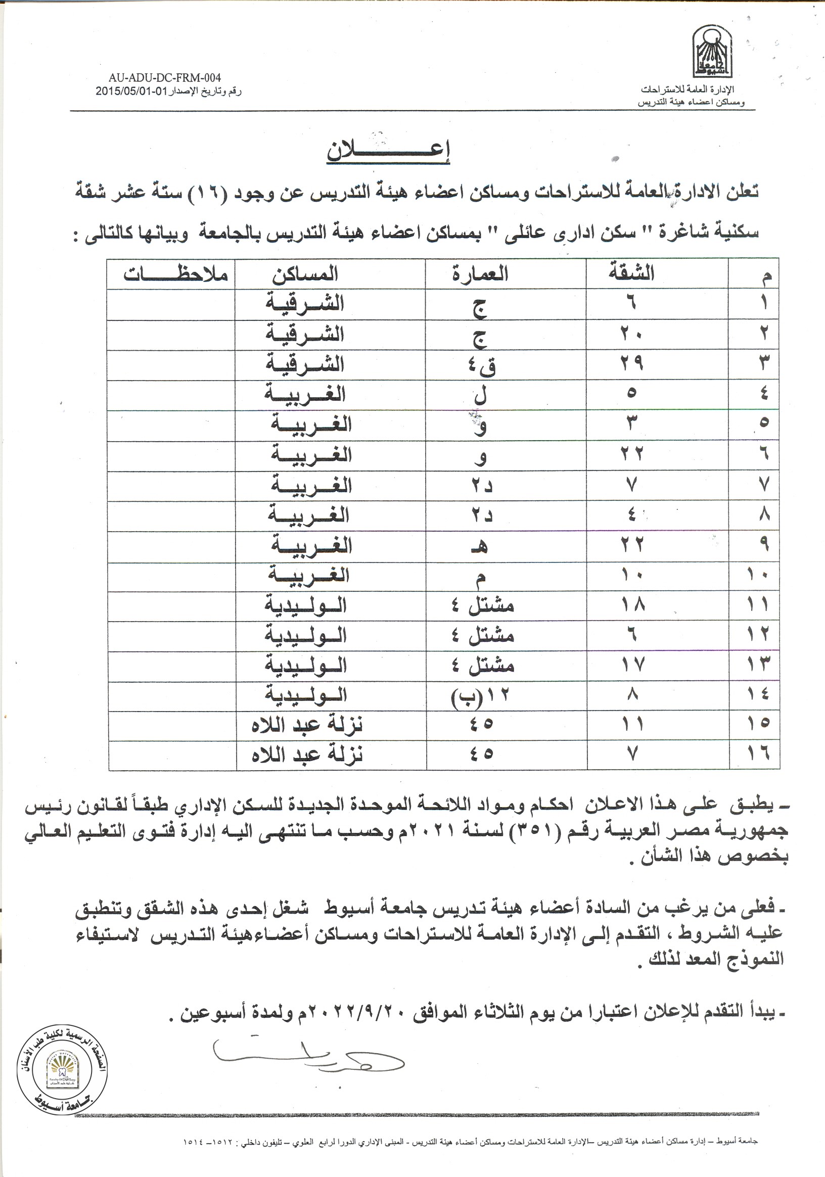
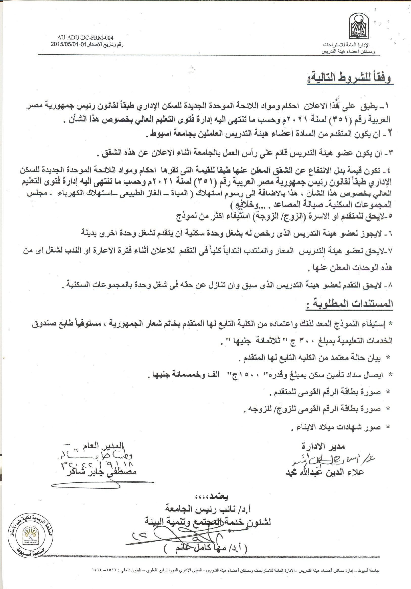
The General Department of Housing announces that there are 16 available departments for staff members and their families.
For more information, kindly read the announcement.
Abstract
BACKGROUND: Piezocision-assisted orthodontics (PAO) is considered one of the modern techniques aiming at reducing the treatment time and overcoming some limitations of orthodontic treatment. The use of piezocision as an adjunct in the treatment of posterior crossbite is limited, so additional research in this area is required.
AIM: To three-dimensionally compare the skeletal and dental effects produced by piezocision-assisted rapid maxillary expansion (PARME) and conventional rapid maxillary expansion (RME) using cone beam computed tomography (CBCT).
MATERIALS AND METHODS: This prospective controlled study comprised 14 consecutive non-syndromic patients with posterior crossbite. In 7 patients (mean age = 16.1 ± 0.3 years), PARME was used to correct the crossbite; whereas in the remaining 7 (mean age = 15.9 ± 0.5 years), RME was done. Cone beam computed tomography (CBCT) scans were performed before expansion (T1) and 3 months later after expansion (T2) to compare the skeletal and dental effects produced by the two expansion techniques. Transverse skeletal, dentolinear, and dentoangular variables at the level of maxillary first and second premolars and maxillary first molars were measured and compared within and between groups using the appropriate statistical test.
RESULTS: For the transverse skeletal variables, PARME showed a non-significant increase; whereas, RME showed a significant increase. Regarding the dentolinear measurements, a significant increase in coronal widths and an insignificant increase in apical widths was seen in PARME, whereas, the RME showed a non-significant increase for both coronal and apical widths. Non-significant decreases (protrusion of teeth) in the dentoangular measurements were seen in both groups. Between-group comparisons showed a non-significant difference except for the dentolinear coronal widths.
CONCLUSION: PARME is effective in treating posterior crossbite. Because of the more dental expansion produced by PARME as compared to the conventional RME, PARME should be limited only to mild or moderate not severe forms of palatal constriction. The available evidence regarding the effectiveness of corticotomy- and/or piezocision-assisted maxillary expansion for correction of posterior crossbite is limited and inadequate.
ABSTRACT
Objective: To elucidate the positional and dimensional temporomandibular joint (TMJ) changes after correction of posterior crossbite in growing patients.
Materials and Methods: A systematic unrestricted search was done in six databases until June 27, 2017. A manual search in the reference lists of the included studies and gray literature was also performed. The eligibility criteria included randomized controlled trials and prospective nonrandomized controlled clinical trials and a sample of growing subjects with posterior crossbite that required maxillary expansion. Risk of bias assessment was performed using Cochrane’s Risk of Bias Tool. The literature search, study inclusion, risk of bias assessment, evaluation of quality of evidence (GRADE), and data extraction were performed by two reviewers independently.
Results: Only two articles were finally eligible to be included in the qualitative analysis. Both studies were RCTs and were assessed as having unclear risk of bias. Meta-analysis was not possible since one study used cone-beam computed tomography as an assessment tool while the other used ultrasonography. One study reported significant reduction in the condylar positional difference between centric and habitual occlusion in the treatment group, while no spontaneous correction of condylar asymmetric position occurred in the control group. The other study reported minor changes of condylar position in both treatment and control groups.
Conclusions: The current available data provide insufficient and weak evidence to form a solid and firm conclusion. There is poor, very low-quality evidence regarding the positional and dimensional effects of posterior crossbite correction on the TMJs. (Angle Orthod. 2018;88:638–648.)
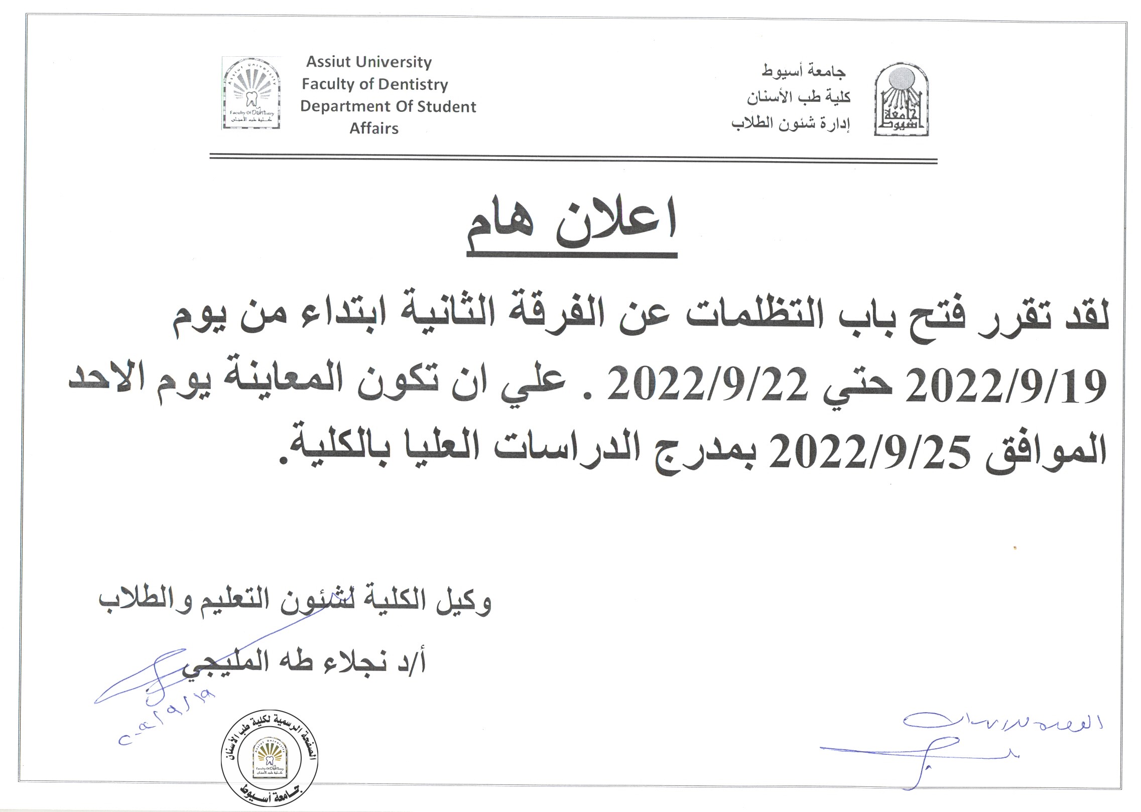
Grievances are available for second year students from 19-9-2022 until 22-9-2022.

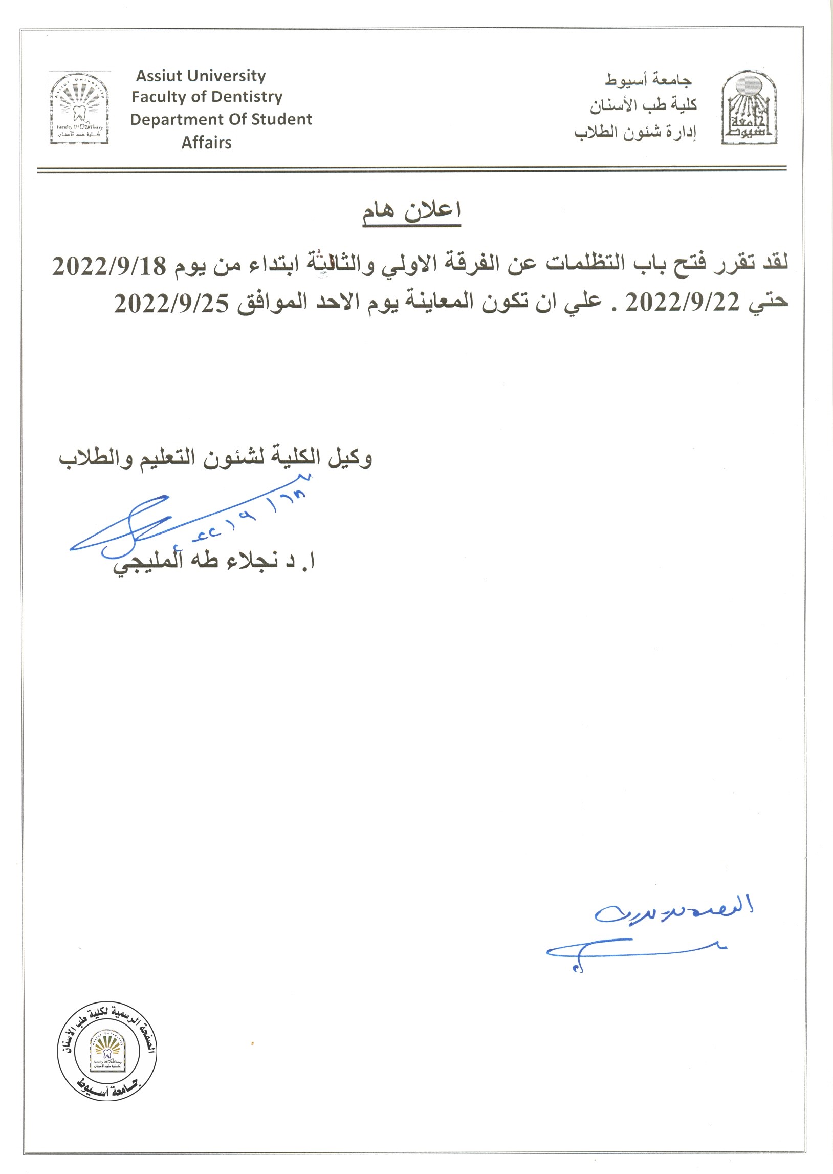
Grievances are available for the first- and third-year students from 18-9-2022 until 22-9-2022.

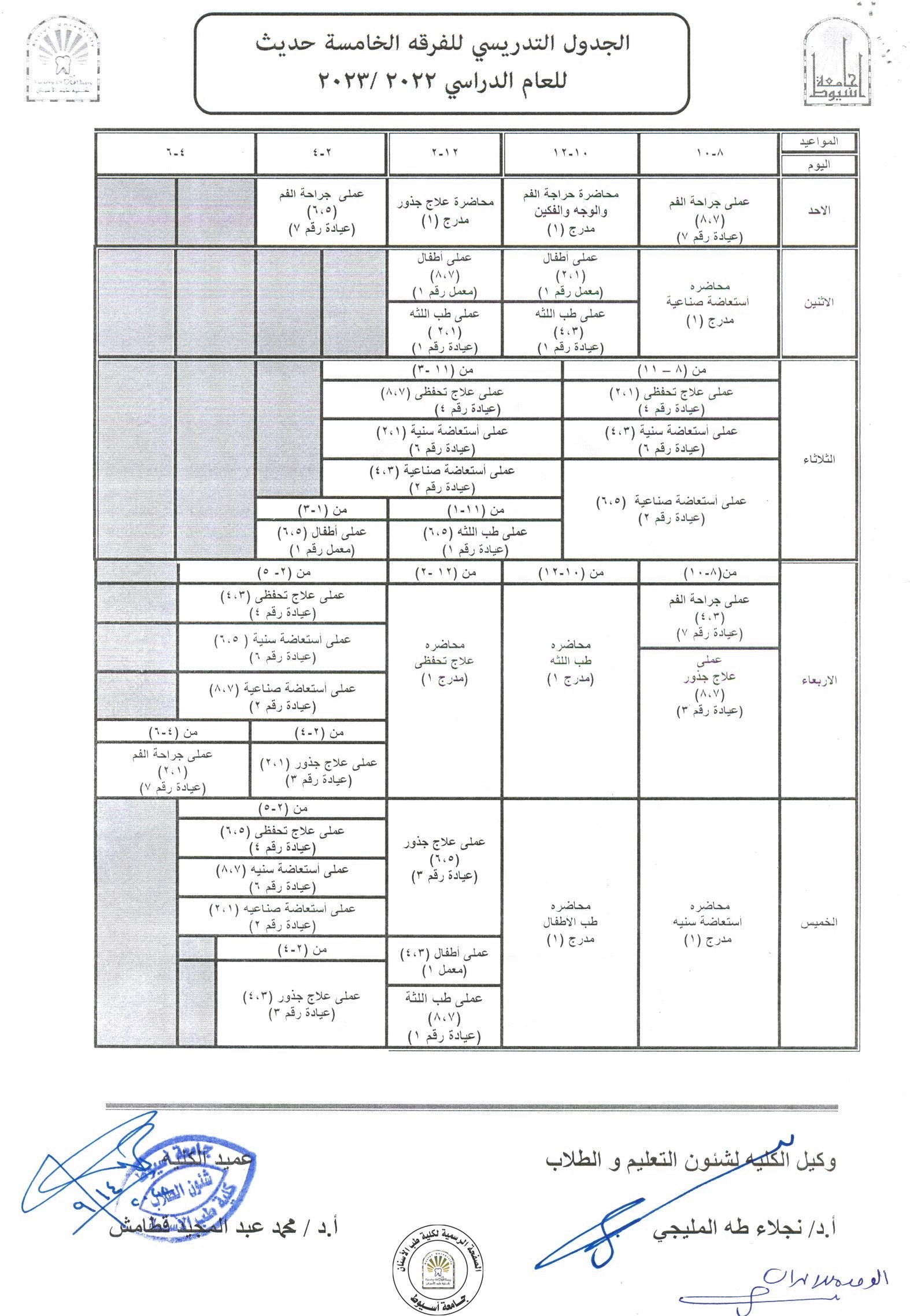
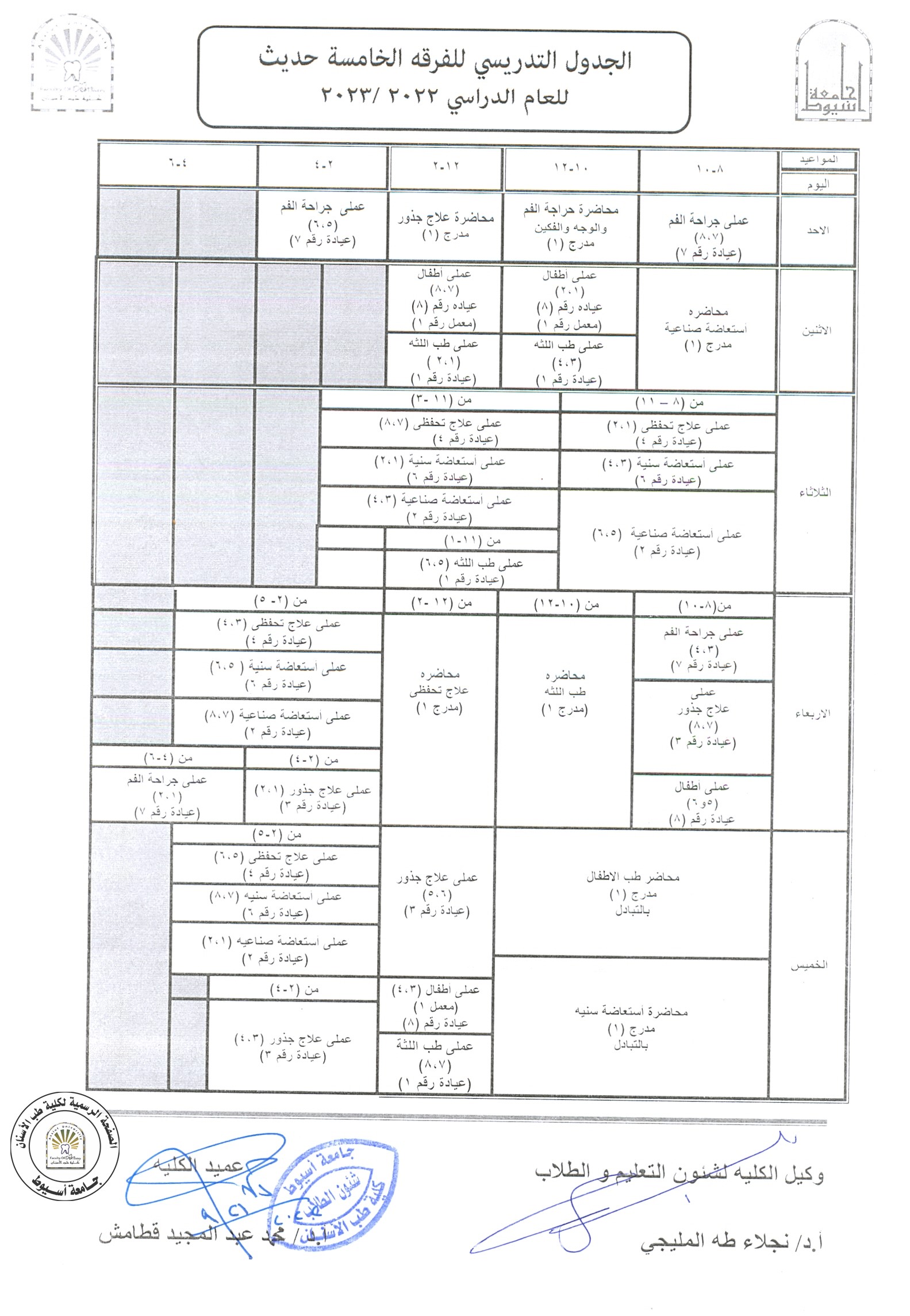
Fifth year timetable for the academic year 2022/2023 is now available.

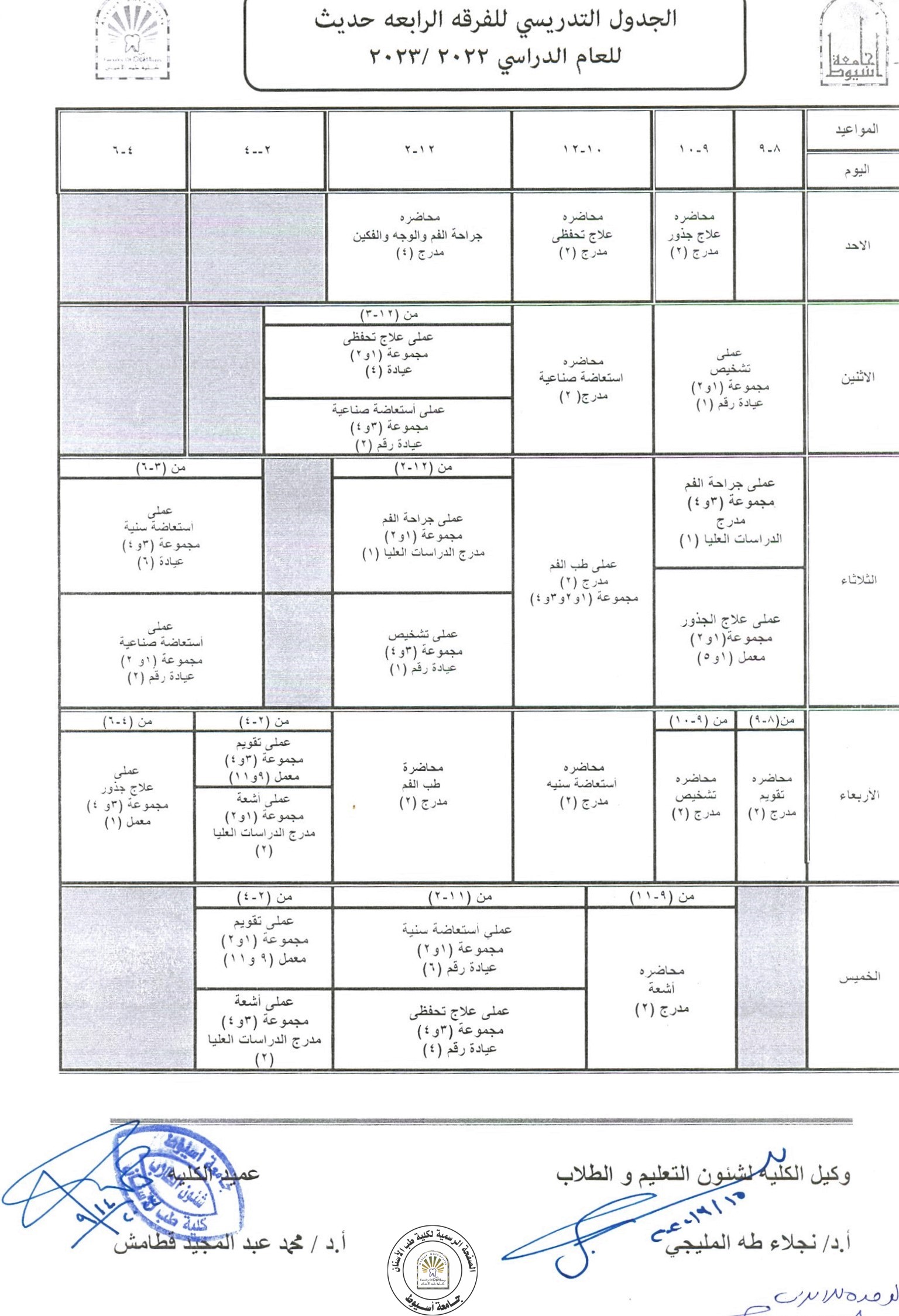
Fourth year timetable for the academic year 2022/2023 is now available.

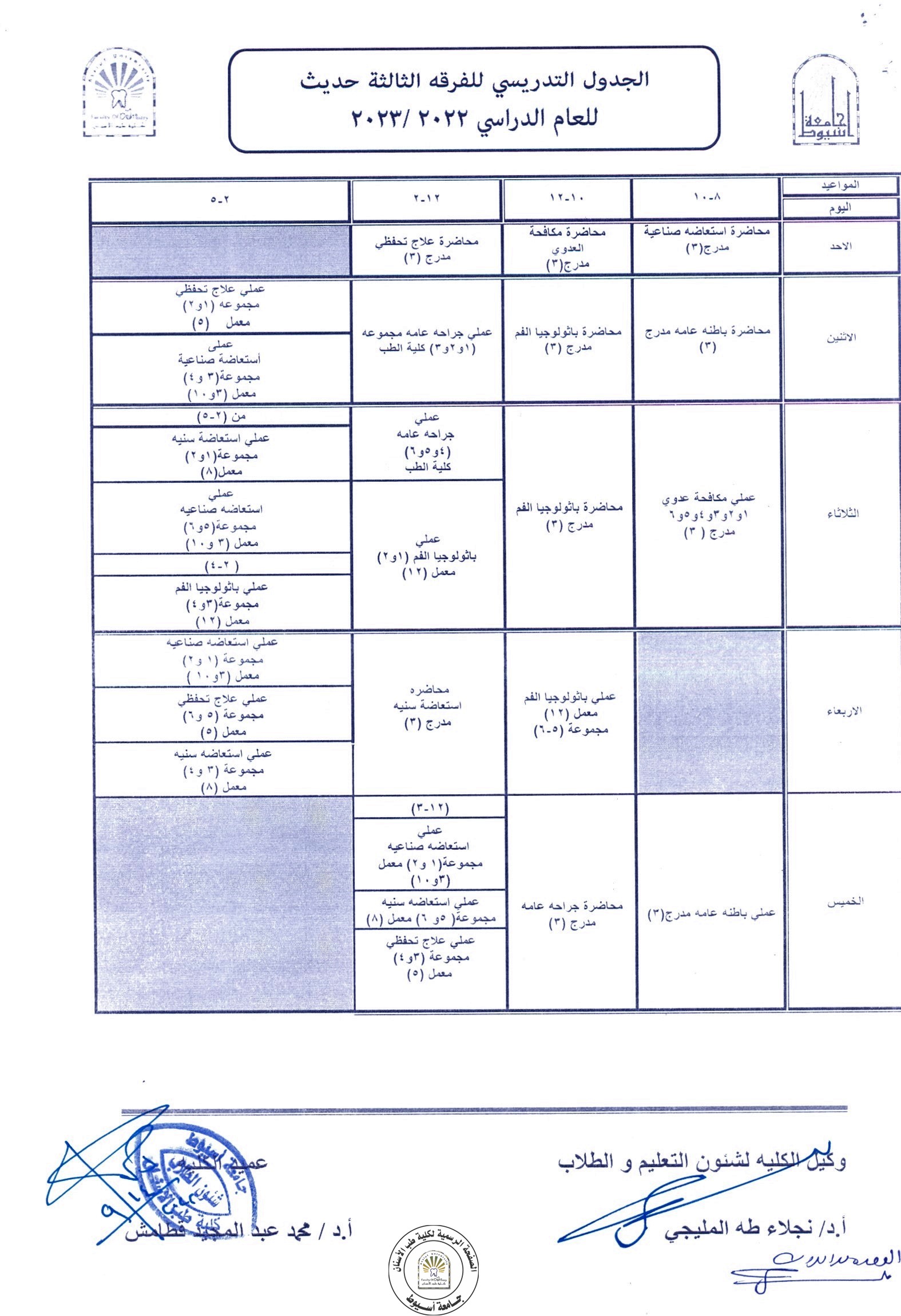
Third Year timetable for the academic year 2022/2023 is now available.

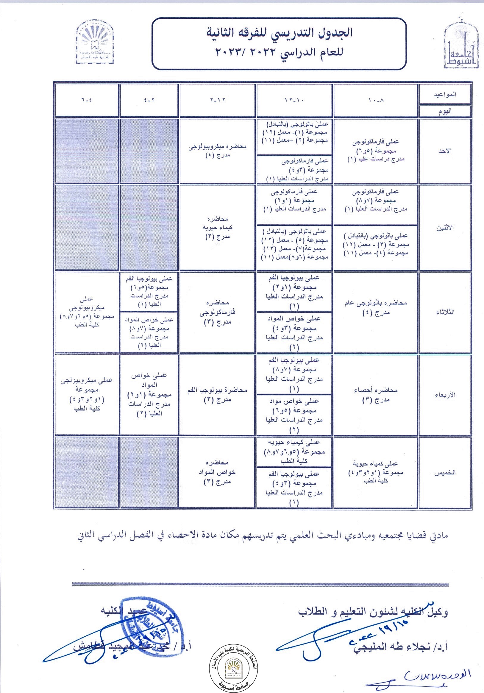
Second Year timetable for the academic year 2022/2023 is now available.