
Ameliorative effects of propolis and wheat germ oil on acute toxoplasmosis in experimentally infected mice are associated with reduction in parasite burden and restoration of histopathological changes in the brain, uterus, and kidney

Research Authors
Ehab Kotb Elmahallawy, Fatma Abo Zakaib Ali, Enrique Raya-Álvarez, Alaa Fehaid, Khaled A. Abd El-Razik, Hassan Ali Mohamed El Fadaly, Manal F. El-Khadragy, Amal S. M. Sayed, Ashraf H. Soror, Alaa S. Alhegaili, Amira A. Saleh, Abdulsalam A. M. Alkhaldi, Ab
Research Date
Research Journal
Frontiers in Veterinary Science

The impact of Pumpkin treatment on TCDD-induced nephrotoxicity in Wister albino rats: biochemical and histopathological studies

Research Abstract

Dynamic economical and industrial expansion lead to environmental contamination and pollution with increasing amounts of harmful agents. Recently, dioxins and dioxin-like compounds (DLCs) have received much attention due to their widespread occurrence, high levels of toxicity, and the significant threat these compounds pose to humans. In this study, we investigated renal toxicity of 2,3,7,8-Tetrachlorodibenzo-p-dioxin (TCDD) and explained the role of pumpkin seed oil in modulating TCDD toxicity in the kidney of Wister albino rats. Thirty male and female Wister rats were divided into three isolated groups ten animal each. Group 1 (G1) served as normal control, group 2 (G2), rats were received single intraperitoneal dose of TCDD (2μg/kg b.w. dissolved in corn oil), while group 3 (G3), rats were treated with oral pumpkin seed oil (PO) (1.5 ml/kg b.w. day after day) after TCDD treatment (same as group II). Blood samples were collected for biochemical analysis and kidney tissue samples for histological examination. our investigation revealed that, TCDD induced significant increase (P<0.01) in urea level and significant increase in creatinine level (P<0.05) in comparison to control group. TCDD treated rats showed series of renal histopathological alterations present mostly in renal vasculature associated with severe degeneration and necrosis of renal tubular epithelium. The previous mentioned lesions were reported much more prominent in renal medulla than in renal cortex. Interestingly, PO treated group showed significant increase (P<0.001) in urea level and significant decrease (P<0.05) in comparison with TCDD group. PO treatment ameliorates these biochemical and pathological changes in renal tissue. In conclusion, exposure to 2, 3, 7, 8-TCDD led to serious toxic effects in renal tissue and treatment with PO could diminish this toxicity to an improved extend.

Research Authors
Hanaa Gamal Abo Bakr Mahmoud A.Z. Sotohy A. SotohyFatma Abo Zakaib Ali
Research Date
Research Department
Research File
Research Member

Splicing of a myosin phosphatase targeting subunit 1 alternative exon is regulated by intronic cis-elements and a novel bipartite exonic enhancer/silencer element

Research Abstract
  1. Isoforms of the smooth muscle myosin phosphatase targeting subunit 1 (MYPT1) are generated by cassette-type alternative splicing of exons. Tissue-specific expression of these isoforms is thought to determine smooth muscle-relaxant properties and unique responses to signaling pathways. We used mini-gene deletion/mutation constructs to identify cis regulators of splicing of the chicken MYPT1 central alternative exon. Comparisons of alternative exon splicing were made between smooth muscle cells of the fast-phasic contractile phenotype (gizzard), in which the central alternative exon is skipped, and slow tonic contractile phenotype (aorta), in which the alternative exon is included. We demonstrate that splicing of the alternative exon requires a cis-enhancer complex in the vicinity of the alternative exon 5'-splice site. This complex consists of two UCUU motifs in an intronic U-rich sequence (putative PTB (polypyrimidine tract binding) or T cell inhibitor of apoptosis-1 binding sites), an intronic 67-nucleotide enhancer that has similarities with the cardiac Troponin T MSE3 enhancer, and a potentially novel exonic splicing enhancer. The exonic enhancer contains the palindromic sequence UCCUACAUCCU present in many other transcripts where alternative splicing of exons occurs, suggesting that it may be more broadly active. The exonic enhancer is adjacent to a potentially novel exonic silencer element that contains a 13-nucleotide imperfect palindromic sequence. This silencer, in conjunction with a distal intronic silencer, is proposed to mediate the silencing of splicing of the MYPT1 central alternative exon in the fast phasic smooth muscle phenotype.
Research Authors
Dirksen WP, Mohamed SA, Fisher SA.
Research Date
Research Member

The impact of Pumpkin treatment on TCDD-induced nephrotoxicity in Wister albino rats: biochemical and histopathological studies

Research Abstract

Dynamic economical and industrial expansion lead to environmental contamination and pollution with increasing amounts of harmful agents. Recently, dioxins and dioxin-like compounds (DLCs) have received much attention due to their widespread occurrence, high levels of toxicity, and the significant threat these compounds pose to humans. In this study, we investigated renal toxicity of 2,3,7,8-Tetrachlorodibenzo-p-dioxin (TCDD) and explained the role of pumpkin seed oil in modulating TCDD toxicity in the kidney of Wister albino rats. Thirty male and female Wister rats were divided into three isolated groups ten animal each. Group 1 (G1) served as normal control, group 2 (G2), rats were received single intraperitoneal dose of TCDD (2μg/kg b.w. dissolved in corn oil), while group 3 (G3), rats were treated with oral pumpkin seed oil (PO) (1.5 ml/kg b.w. day after day) after TCDD treatment (same as group II). Blood samples were collected for biochemical analysis and kidney tissue samples for histological examination. our investigation revealed that, TCDD induced significant increase (P<0.01) in urea level and significant increase in creatinine level (P<0.05) in comparison to control group. TCDD treated rats showed series of renal histopathological alterations present mostly in renal vasculature associated with severe degeneration and necrosis of renal tubular epithelium. The previous mentioned lesions were reported much more prominent in renal medulla than in renal cortex. Interestingly, PO treated group showed significant increase (P<0.001) in urea level and significant decrease (P<0.05) in comparison with TCDD group. PO treatment ameliorates these biochemical and pathological changes in renal tissue. In conclusion, exposure to 2, 3, 7, 8-TCDD led to serious toxic effects in renal tissue and treatment with PO could diminish this toxicity to an improved extend.

Research Authors
Hanaa Gamal Abo Bakr Mahmoud A.Z. Sotohy A. Sotohy Fatma Abo Zakaib Ali
Research Date
Research Department
Research Member

Histomorphmetric studies on the protective role of Maca (Lepidium meyenii) on New Zealand White Rabbits prostate gland under oxidative stress

Research Abstract

The prostate gland considers the most important gland among rabbits' accessory genital glands and secretes many factors essential for sperm function. This study aims to show the impact of Maca as an antioxidant on the prostate gland's histomorphology and its capacity to reduce the effects of oxidative stress. Twenty-four healthy New Zealand white male rabbits, 6-8 months divided into 4 groups.1st group (control group), 2nd group (Maca), 3rd group (H2O2), 4th group (Maca+ H2O2). To evaluate these effects we used histomorphometrical studies. In control and Maca groups, acini were lined by simple columnar epithelium and showed normal interstitium. However, in H2O2 treated rabbits, we observed cuboidal epithelium and epithelium height decreased significantly compared to other groups, and interstitium showed fibrosis. Also in H2O2 treated rabbits, we observed mild degenerative changes in the epithelial lining the acini, with karyolitic nucleus and vacuolated cytoplasm. In concern to acini length, we observed that the secretory units in Maca groups was overcrowd due to increasing acini length. However, in H2O2 exposed rabbits the diameter of the glandular portion and the length of the acini decreased significantly compared to other groups. Moreover in H2O2 treated rabbits we demonstrated other changes as congested and hyperemic blood vessels and muscular layer appeared disorganized and degenerated. In Maca+ H2O2 (H+M) treated rabbits the epithelium reverted to simple columnar and the secretory activity increased compared to H2O2 group. In conclusion, we can ensure the effect of Maca as an anti-oxidant and its ameliorative effect on oxidative stressed rabbits.

Research Authors
Asmaa Nabil; Fatma El-Zahraa A. Mustafa; Enas A. Abdelhefez; Mostafa Galal Abdelfattah; M. A. M. Sayed; Manal T Hussein
Research Date
Research Journal
svu, international veterinary journal
Research Member
Research Pages
14
Research Publisher
asmaa nabil
Research Rank
2.5
Research Vol
6
Research Year
2023

Histomorphmetric studies on the protective role of Maca (Lepidium meyenii) on New Zealand White Rabbits prostate gland under oxidative stress

Research Abstract

The prostate gland considers the most important gland among rabbits' accessory genital glands and secretes many factors essential for sperm function. This study aims to show the impact of Maca as an antioxidant on the prostate gland's histomorphology and its capacity to reduce the effects of oxidative stress. Twenty-four healthy New Zealand white male rabbits, 6-8 months divided into 4 groups.1st group (control group), 2nd group (Maca), 3rd group (H2O2), 4th group (Maca+ H2O2). To evaluate these effects we used histomorphometrical studies. In control and Maca groups, acini were lined by simple columnar epithelium and showed normal interstitium. However, in H2O2 treated rabbits, we observed cuboidal epithelium and epithelium height decreased significantly compared to other groups, and interstitium showed fibrosis. Also in H2O2 treated rabbits, we observed mild degenerative changes in the epithelial lining the acini, with karyolitic nucleus and vacuolated cytoplasm. In concern to acini length, we observed that the secretory units in Maca groups was overcrowd due to increasing acini length. However, in H2O2 exposed rabbits the diameter of the glandular portion and the length of the acini decreased significantly compared to other groups. Moreover in H2O2 treated rabbits we demonstrated other changes as congested and hyperemic blood vessels and muscular layer appeared disorganized and degenerated. In Maca+ H2O2 (H+M) treated rabbits the epithelium reverted to simple columnar and the secretory activity increased compared to H2O2 group. In conclusion, we can ensure the effect of Maca as an anti-oxidant and its ameliorative effect on oxidative stressed rabbits.

Research Authors
Asmaa Nabil; Fatma El-Zahraa A. Mustafa; Enas A. Abdelhefez; Mostafa Galal Abdelfattah; M. A. M. Sayed; Manal T Hussein
Research Date
Research Member

The President of Assiut University renews the appointment of Professor Dr. Madiha Darwish - Dean of the College - as General Coordinator of Student Activities at Assiut University

نقلا عن الصفحة الرسمية لجامعة اسيوط

h


2

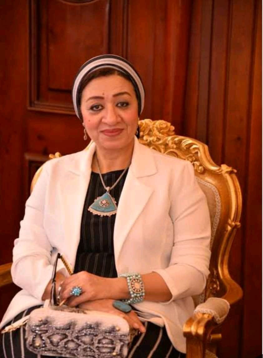
رئيس جامعة أسيوط يجدد تكليف الدكتورة مديحة درويش منسقاً عاماً للأنشطة الطلابية بجامعة أسيوط 

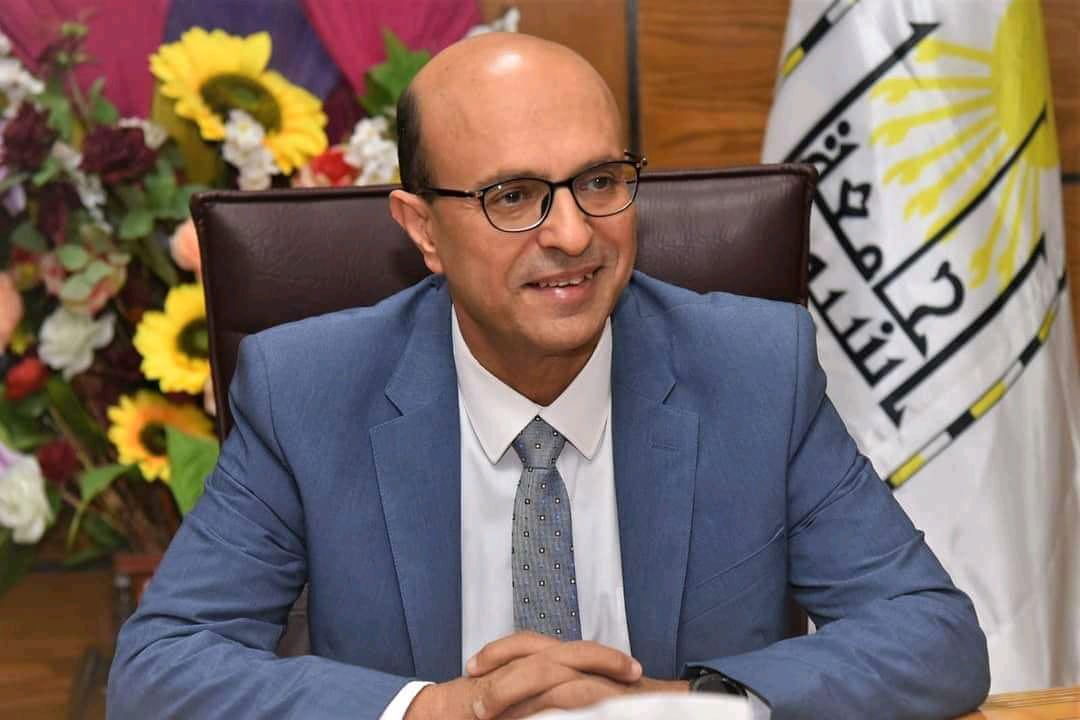
 أصدر الدكتور أحمد المنشاوي رئيس جامعة أسيوط، قراراً بتجديد تكليف الدكتورة مديحة حسني أحمد درويش عميد كلية الطب البيطري؛ منسقاً عاماً للأنشطة الطلابية بالجامعة، وذلك لمدة عام.

   وهنأ الدكتور المنشاوى؛ الدكتورة مديحة درويش علي تجديد تكليفها لهذا المنصب المهم، موجهاً بمواصلة دعم ورعاية كافة الأنشطة الطلابية، والمشاركة في مختلف المحافل والمسابقات، على المستوى المحلي والاقليمي والدولي، مشيرا إلى أهمية اكتشاف المواهب الشابة واحتضانها ودعم قدراتها، وفتح مسارات جديدة للأنشطة الطلابية، وذلك في إطار من المعرفة، والفكر السليم، ودعم قيم الوﻻء، والانتماء ﻟلوطن.

   جدير بالذكر، أن الدكتورة مديحة درويش شغلت عدداً من المناصب الأكاديمية، بكلية الطب البيطري، من بينها: رئيساً لقسم السلوكيات ورعاية الحيوان والدواجن، ورئيساً لقسم طب الأحياء المائية ورعايتها، ووكيلاً للكلية لشئون الدراسات العليا والبحوث، ثم عميداً للكلية، ومستشاراً للجنة العليا للأنشطة الطلابية، ومنسقاً عاماً للأنشطة الطلابية بالجامعة.
#إعلام_جامعة_أسيوط

رابط الخبر 

https://www.facebook.com/100068448236848/posts/760677992890468/?app=fbl

Exploring Cellular Dynamics in the Goldfish Bulbus Arteriosus: A Multifaceted Perspective

Research Abstract

The bulbus arteriosus of goldfish, Carassius auratus, possesses unique structural features.
The wall of the bulbus arteriosus is exceptionally thick, with an inner surface characterized by
longitudinally arranged finger-like ridges, resulting in an uneven luminal appearance. These ridges
are covered by endocardium and encased in an amorphous extracellular matrix. The inner surface
of the bulbus arteriosus also contains rodlet cells at different developmental stages, often clustered
beneath the endothelium lining the bulbar lumen. Ruptured rodlet cells release their contents via
a holocrine secretion process. The high abundance of rodlet cells in the bulbus arteriosus suggests
that this is the site of origin for these cells. Within the middle layer of the bulbus arteriosus, smooth
muscle cells, branched telocytes (TCs), and collagen bundles coexist. TCs and their telopodes form
complex connections within a dense collagen matrix, extending to rodlet cells and macrophages.
Moreover, the endothelium makes direct contact with telopodes. The endocardial cells within the
bulbus arteriosus display irregular, stellate shapes and numerous cell processes that establish direct
contact with TCs. TEM reveals that they contain moderately dense bodies and membrane-bound
vacuoles, suggesting a secretory activity. TCs exhibit robust secretory activity, evident from their
telopodes containing numerous secretory vesicles. Furthermore, TCs release excretory vesicles
containing bioactive molecules into the extracellular matrix, which strengthens evidence for telocytes
as promising candidates for cellular therapies and regeneration in various heart pathologies.
 

Research Authors
3. Mokhtar, D.M.; Abd-Elhafez, E.A.; Albano, M.; Zaccone, G.; Hussein, M.T.
Research Date
Research Journal
Fishes
Research Publisher
MDPI
Research Rank
Q2
Research Website
https://doi.org/10.3390/fishes9060203
Research Year
2024

Neuron mapping in the Molly fish optic tectum: An emphasis on the adult neurogenesis process

Research Abstract

Teleost fish exhibit the most pronounced and widespread adult neurogenesis.
Recently, functional development and the fate of newborn neurons have been
reported in the optic tectum (OT) of fish. To determine the role of neurogenesis in
the OT, this study used histological, immunohistochemical, and electron microscopic
investigations on 18 adult Molly fish specimens (Poecilia sphenops). The OT of the
Molly fish was a bilateral lobed structure located in the dorsal part of the mesencephalon. It exhibited a laminated structure made up of alternating fiber and cellular layers,
which were organized into six main layers. The stratum opticum (SO) was supplied by
optic nerve fibers, in which the neuropil was the main component. Radial bipolar neurons that possessed bundles of microtubules were observed in the stratum fibrosum
et griseum superficiale (SFGS). Furthermore, oligodendrocytes with their processes
wrapped around the nerve fibers could be observed. The stratum album centrale
(SAC) consisted mainly of the axons of the stratum griseum centrale (SGC) and the
large tectal, pyriform, and horizontal neurons. The neuronal cells of the SO and large
tectal cells of the SAC expressed autophagy-related protein-5 (APG5). Interleukin-1β
(IL-1β) was expressed in both neurons and glia cells of SGC. Additionally, inducible
nitric oxide synthase (iNOS) was expressed in the neuropil of the SAC synaptic layer
and granule cells of the stratum periventriculare (SPV). Also, transforming growth factor beta (TGF-β), SRY-box transcription factor 9 (SOX9), and myostatin were clearly
expressed in the proliferative neurons. In all strata, S100 protein and Oligodendrocyte
Lineage Transcription Factor 2 (Olig2) were expressed by microglia, oligodendrocytes,
and astrocytes. In conclusion, it was possible to identify different varieties of neurons
in the optic tectum, each with a distinct role. The existence of astrocytes, proliferative
neurons, and stem cells highlights the regenerative capacity of OT.
 

Research Authors
Hussein, M. T., Sayed, R. K. A., & Mokhtar, D. M.
Research Date
Research Journal
Microscopy Research and technique
Research Publisher
Wiely Online Library
Research Rank
Q1
Research Website
https://doi.org/10. 1002/jemt.24617
Research Year
2024
Subscribe to