Opening the door for candidacy for the student union elections at Assiut University for the academic year 2022/2021
International Conference in Dubai
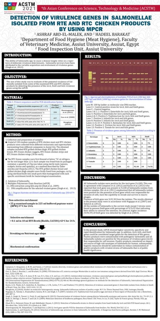
dr. Ashraf Abdel Malik, professor of health control over meat, participated in an international conference in Dubai, with a research in the form of a poster presentation
And there is a competition for the participants. Support and support is required for him by watching, like and share, because he represents the college and the university in this international conference.
Saturday 11/20 - 6 o'clock Cairo time
Link to Channel: https://www.youtube.com/c/TheACSEofficial