 Do you have any questions?
 Congratulations - to the doctor - Mahmoud Saad - resident doctor in the Department of Orthopedics
ا. د/علاء عطية عميد كلية الطب ورئيس مجلس إدارة المستشفيات الجا
ا. د/يوسف صالح وكيل الكلية لشئون الدراسات العليا والبحوث
يهنئون
ط/ محمود سعد
طبيب مقيم بقسم جراحة العظام
وذلك لدوره في نشر ورقة بحثية في مجلة British Medical Journal (open journal)
حيث عمل عام ٢٠٢٠ كمنسق قومي للفرق المصرية المشاركة في دراسة دولية متعددة المراكز تهدف إلى تقييم تداعيات جائحة كوفيد-١٩ على طرق علاج حالات سرطان الأطفال.
الأطفال المصابون بالسرطان في الدول منخفضة ومتوسطة الدخل يعانون من احتمالية وفاة أكثر بأربعة أضعاف من أقرانهم في الدول ذات الدخل المرتفع، ويرجع ذلك إلى عدد من الأسباب منها أن الدول مرتفعة الدخل تتمتع بالتشخيص المبكر، والوعي المرتفع، وتوافر وسائل العلاج الحديثة.
وقد أثبتت هذة الدراسة أن الأطفال المصابين بالسرطان في الدول منخفضة الدخل أكثر عرضة لتغيير خطة العلاج المتبعة لهم في ظل تفشي جائحة كوفيد-١٩ بنسبة تتجاوز ٨٠% من أقرانهم في الدول مرتفعة الدخل ممن يغيرون خطة العلاج مما يؤدي إلى تضاعف أكبر في احتمالية الوفاة نتيجة السرطان.
نسأل الله أن يخفف عن أولئك الأطفال معاناة المرض، وأن يخفف عن ذويهم معاناة الفقر، وأن يحفظ بلادنا من الأوبئة والأسقام.
تتميز الدراسة بكونها الأولى من نوعها بهذا الحجم على مستوى العالم بمشاركة ٩١ مستشفى ومركز لعلاج الأورام من ٣٩ دولة مختلفة بتنسيق عالمي من
Global Health Research Group on Children’s Non-Communicable Diseases
Collaborative.
رابط البحث لمن يريد المزيد من التفاصيل:
https://bmjopen.bmj.com/content/12/4/e054690


The Faculty of Medicine has reached the 451-500 rank globally in the results of the QS English classification of subjects for the year 2.22.
ا. د/علاء عطية عميد كلية الطب ورئيس مجلس إدارة المستشفيات الجامعية
ا. د/ اماني عمر وكيل الكلية لشئون التعليم والطلاب
ا. د/ يوسف صالح وكيل الكلية لشئون التعليم والطلاب
ا. د/سعد زكي محمود وكيل الكلية لشئون خدمة المجتمع وتنمية البيئة
يهنئون
ا. د/ طارق الجمال
رئيس الجامعة
ا. د/ احمد المنشاوي
نائب رئيس الجامعة لشئون الدراسات العليا والبحوث
لوصول كلية الطب الي المرتبه ٤٥١ -٥٠٠ عالميا في نتائج تصنيف QS الانجليزي للموضوعات لعام ٢.٢٢.
هذا التصنيف والصادر من هيئة كواكواريلي سيموندس الانجليزيه يختص بالإضافة الي البحث العلمي والنشر، بسمعة الجامعات في الهيئات الأكاديمية الدولية بالاضافه الي سمعة الجامعة لدي خريجيها. وكذلك تشمل مؤشرات التقيم عدد الطلاب وأعضاء هيئة التدريس الأجانب بالجامعة.
والشكر موصول للسادة..
ا. د/عمر ممدوح شعبان مدير مكتب التصنيف الدولى بجامعة أسيوط
وا. د/ محمد حمد نائب مدير مكتب التصنيف الدولي للجامعة
لدورهم في هذا الانجاز ووضع الخط الاستراتيجية للنهوض بتصنيف جامعة اسيوط العريقة.
Congratulations - to Dr. Hilal Fouad Hetah
أ. د/علاء عطية عميد كلية الطب ورئيس مجلس إدارة المستشفيات الجامعية.
أ. د/ اماني عمر وكيل الكلية لشئون التعليم والطلاب
أ. د/ يوسف صالح وكيل الكلية لشئون الدراسات العليا والبحوث
أ. د/ سعد زكي محمود وكيل الكلية لشئون خدمة المجتمع وتنمية البيئة.
يهنئون
د/ هلال فؤاد حته
أستاذ مساعد الميكروبيولوجي و المناعه الطبيه
بفوزه بجائزة الدولة التشجيعيه للعلوم الطبيه لعام ٢٠٢١ التي تمنحها أكاديمية البحث العلمي والتكنولوجيا سنويًا.
Urology and Kidney Hospital celebrated the completion of the fiftieth process of kidney transplantation
تحت رعاية
ا. د/ طارق الجمال رئيس الجامعة
ا. د/علاء عطية عميد كلية الطب ورئيس مجلس إدارة المستشفيات الجامعية
و ا. د/ ايهاب فوزي المدير التنفيذي للمستشفيات الجامعية.
اتمام ٥٠ عملية زراعة كلي بجامعة اسيوط
احتفلت مستشفي جراحة المسالك البولية والكلي بإتمام العملية الخمسون زراعة كلى بواسطة الفريق الطبي المختص بعمليات الزرع والمكون من ٥٠ فرد من الاطقم الطبية من الاقسام المختلفة من اساتذة واطباء وتمريض بقيادة ا. د/هشام مختار حمودة الأستاذ المتفرغ بقسم جراحة الكلى والمسالك بالمستشفى ومدير وحدة زراعة الكلى بالمستشفى.
وقريبا بدء الاستعدادات لتنفيذ برنامج زراعة الكلى للأطفال عقب الزيارة العلمية التي قام بها ا. د/ هشام مختار حمودة أستاذ جراحة المسالك البولية ومدير برنامج زراعه الكلى وعضو اللجنة العليا لزراعه الأعضاء وذلك بعد الاضطلاع على احدث التقنيات والبروتوكولات المستخدمة فى زراعه الكلى للأطفال بمستشفى الأطفال بجامعه نوتنجهام بالمملكة المتحدة تمهيدا لتطبيقها فى جامعة أسيوط
ويعد انفراد جامعة أسيوط بذلك النوع من عمل زرع للكلى على مستوى صعيد مصر، تزامناً هذا الاهتمام مع توجيهات القيادة السياسية للتوسع فى زراعه الكلى كعلاج ناجز لمرض الفشل الكلوي.
Forum for self-seminars at the university level
تحت رعاية
ا. د/ طارق الجمال رئيس جامعة أسيوط.
ا. د/ شحاته غريب نائب رئيس الجامعة لشئون التعليم والطلاب.
ا. د/ علاء عطية عميد كلية الطب ورئيس مجلس إدارة المستشفيات الجامعية
ا. د/ اماني عمر وكيل الكلية لشئون التعليم والطلاب
قام فريق مكون من 6 طلاب من كلية الطب البشري بالمشاركة في
والمدن الجامعية
بموضوع بعنوان
" فيروس سي ودور مصر في القضاء عليه"
وقد اشادت لجنة التحكيم بعمل الطلاب، وأدائهم المميز جدا والمشرف في العرض الذى يليق باسم كلية الطب، وخصوصا الطالبة إسراء عصام.
والطلاب المشاركون هم :
١_ ياسر اشرف جمال الفرقة الثالثة
٢_ احمد محمد الصاوي الفرقة الثالثة
٣_ محمد احمد فواد الفرقة الأولى
٤_ هند ناجح عبدالحكيم الفرقة الثالثة
٥_ علا احمد عبدالرحمن هارون الفرقة الثالثة
٦_ اسراء عصام السيد محمد الفرقة السادسة
والشكر موصول للسادة:
الاستاذ الدكتور/ على محمد عبد الرحيم مستشار إعداد القادة.
الأستاذ/ عز المنصورى مدير الإدارة العامة لرعاية الشباب المركزية.
الأستاذ/ أشرف البلك مدير إدارة إعداد القادة.
مشرف النشاط الاستاذ/ علي حامد القرن
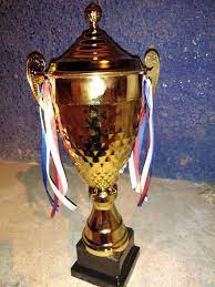
Handing a cup - the first Ramadan session for departments and departments - Faculty of Medicine
تحت رعاية
أ.د/ طارق الجمال - رئيس الجامعة
ا. د/علاء عطية عميد كلية الطب ورئيس مجلس إدارة المستشفيات الجامعية
ا. د/ يوسف صالح وكيل الكلية لشئون الدراسات العليا والبحوث
تسليم كأس المركز الاول
فريق قسم جراحة المسالك البولية
كأس المركز الثانى
فريق الامتياز
وشهادات التقدير للسادة المشاركين
في
الدورة الرمضانية الأولى لاقسام و إدارات
كلية الطب
بملاعب كلية التربية الرياضية بجوار كلية فنون جميلة
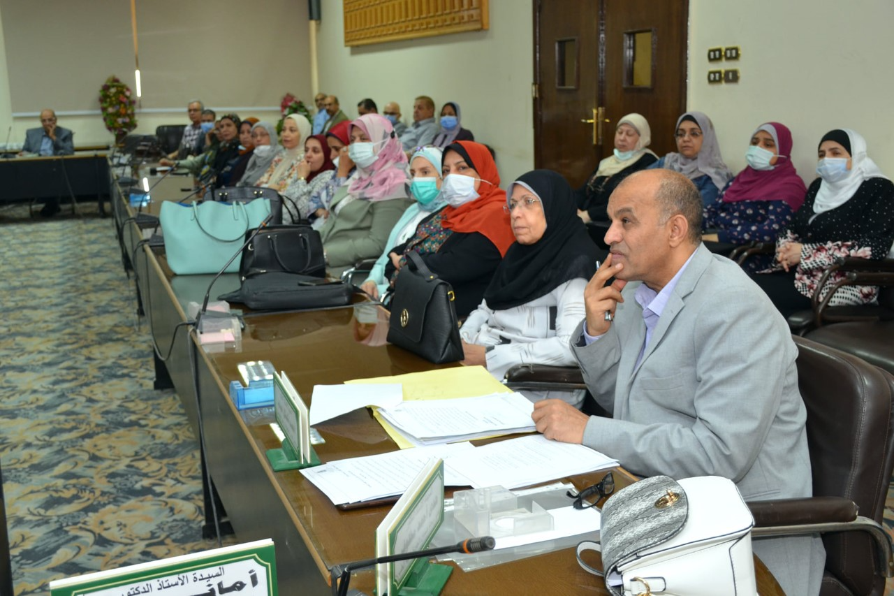
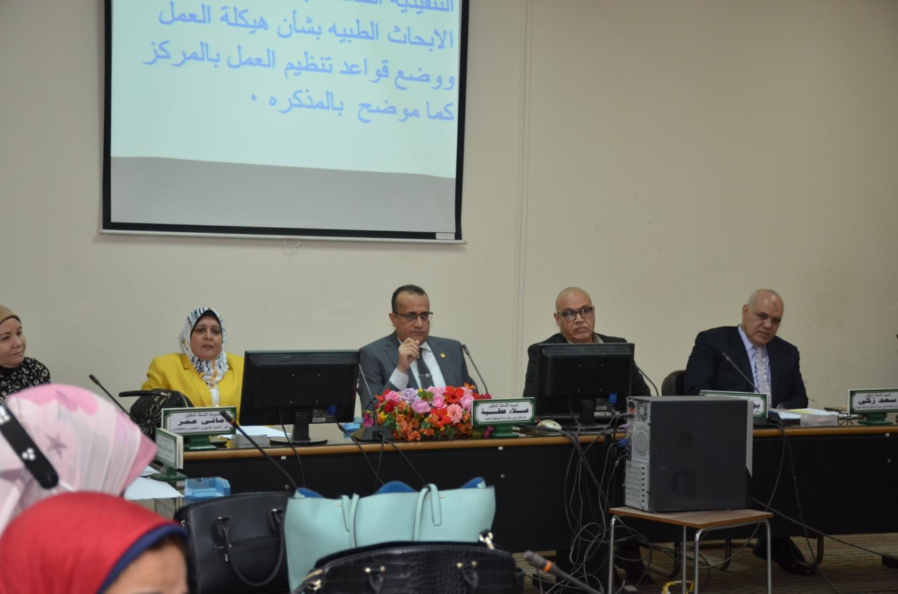
College Council meeting - for the month of April 2022 AD
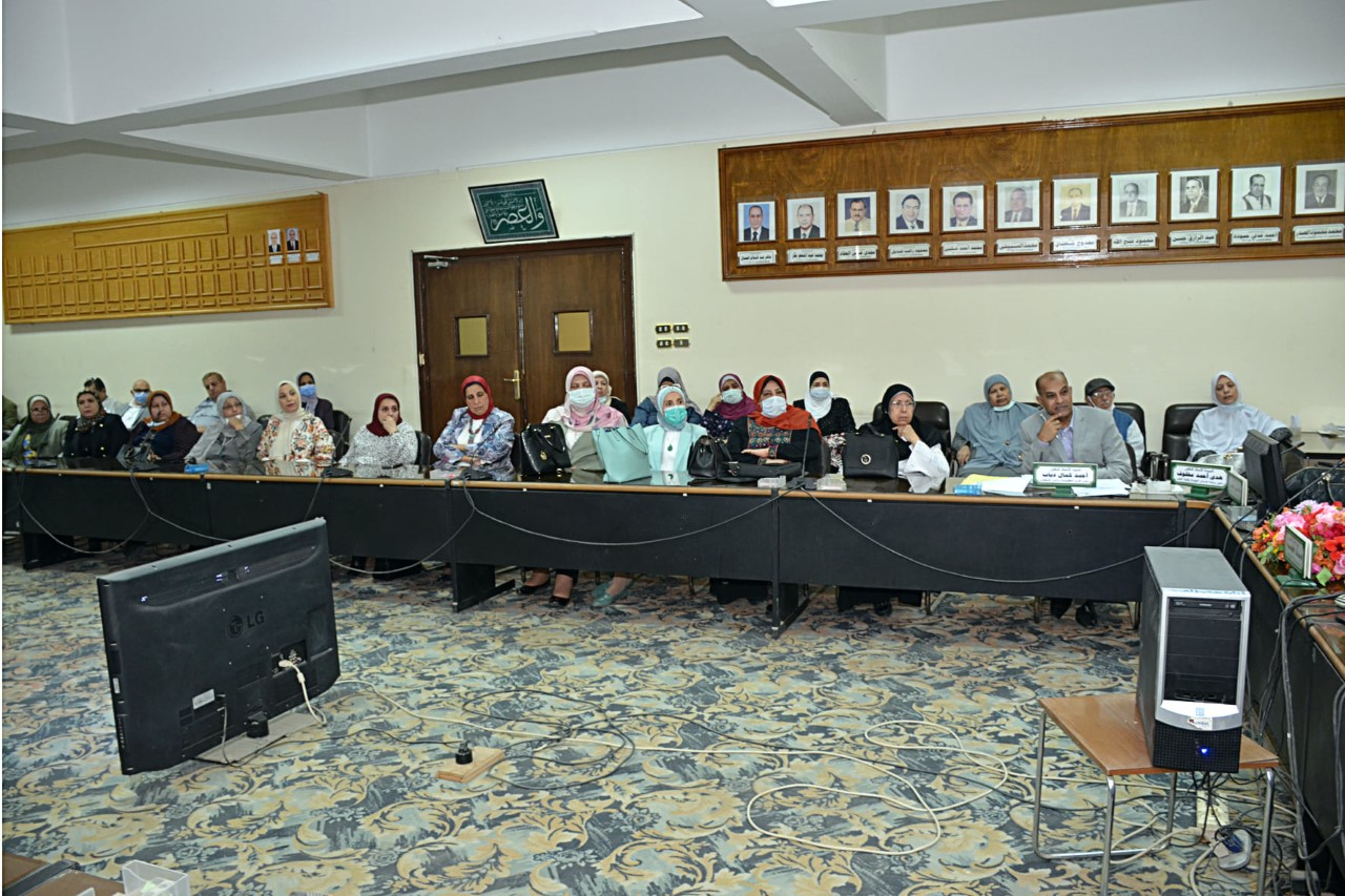
إنعقاد مجلس الكلية
يوم الاحد ٢٠٢٢/٤/٢٤
بحضور
أ. د/علاء عطية عميد كلية الطب ورئيس مجلس إدارة المستشفيات الجامعية.
أ. د/ اماني عمر وكيل الكلية لشئون التعليم والطلاب
أ. د/ يوسف صالح وكيل الكلية لشئون الدراسات العليا والبحوث
أ. د/ سعد زكي محمود وكيل الكلية لشئون خدمة المجتمع وتنمية البيئة.
ا. د/ احمد دياب استاذ الطفيليات وامين المجلس
ا. د/ هدي مخلوف مدير وحدة ضمان الجودة
وبحضور السادة رؤساء الأقسام واعضاء هيئة التدريس وناقش المجلس سير العمل بالكلية ومشروعاتها
Fifth Scientific Conference Orthopedic Department
تحت رعاية
أ. د/ طارق الجمال رئيس الجامعة
أ. د/ احمد المنشاوي نائب رئيس الجامعة لشئون الدراسات العليا والبحوث
أ. د/ مها كامل غانم نائب رئيس الجامعة لشئون خدمة المجتمع وتنمية البيئة
ا. د/ علاء عطية عميد كلية الطب ورئيس مجلس إدارة المستشفيات الجامعية.
أ . د إيهاب فوزى المدير التنفيذى للمستشفيات الجامعية
. د / محمد الشرقاوي رئيس القسم ورئيس المؤتمر
ا. د/ محمد مهران سكرتير المؤتمر
CD4+ CD25+ CD127-Foxp3+ and CD8+ CD28-Tregs in Renal Transplant Recipients: Phenotypic Patterns, Association With Immunosuppressive Drugs, and Interaction With Effector CD8+ T
Research Abstract
Gaps still exist regarding knowledge on regulatory cells in transplant recipients. We studied the phenotypic patterns of CD4+, CD8+CD28- Tregs, and CD19+IL-10+ Bregs in the blood of healthy controls (HC), end-stage kidney disease patients (ESKD), early and late stable renal transplant recipients (Tx), and transplant recipients with steroid-treated acute cellular rejection 1 week–3 months after successful treatment. We also investigated the relationship between immunosuppressive drugs and the aforementioned regulatory cells in transplant recipients. Methods We recruited 32 HC, 83 HD, 51 early Tx, 95 late Tx, and 9 transplant patients with a recent steroid-treated acute cellular rejection. Besides CD19+IL-10+ Bregs, we analyzed absolute and relative frequencies of CD4+CD25+CD127-Foxp3+ Tregs and CD8+CD28- Tregs and their expression of IL-10, TGF-ß, IFN-g, and Helios. Results We found a negative correlation between absolute CD4+CD25+CD127-Foxp3+ Treg and relative CD19+IL-10+ Breg frequencies in early Tx recipients (r=-0.433, p=0.015, n=31). In that group, absolute CD4+CD25+CD127-Foxp3+ Tregs were negatively associated with steroid dose and tacrolimus trough levels (r=-0.377, p = 0.021, n=37; r=-0.43, p=0.033, n=25, respectively), opposite to IL-10+ Bregs, whose frequency apparently was not negatively affected by potent immunosuppression early posttransplant. We found also lower CD4+CD25+CD127-Foxp3+ Tregs in patients treated with basiliximab or rATG as compared with ESKD patients (p=0.001 and p <0.001, respectively). No difference in absolute IL-10+ Bregs could be detected among these 3 patient …
Research Date
Research Member