 Do you have any questions?
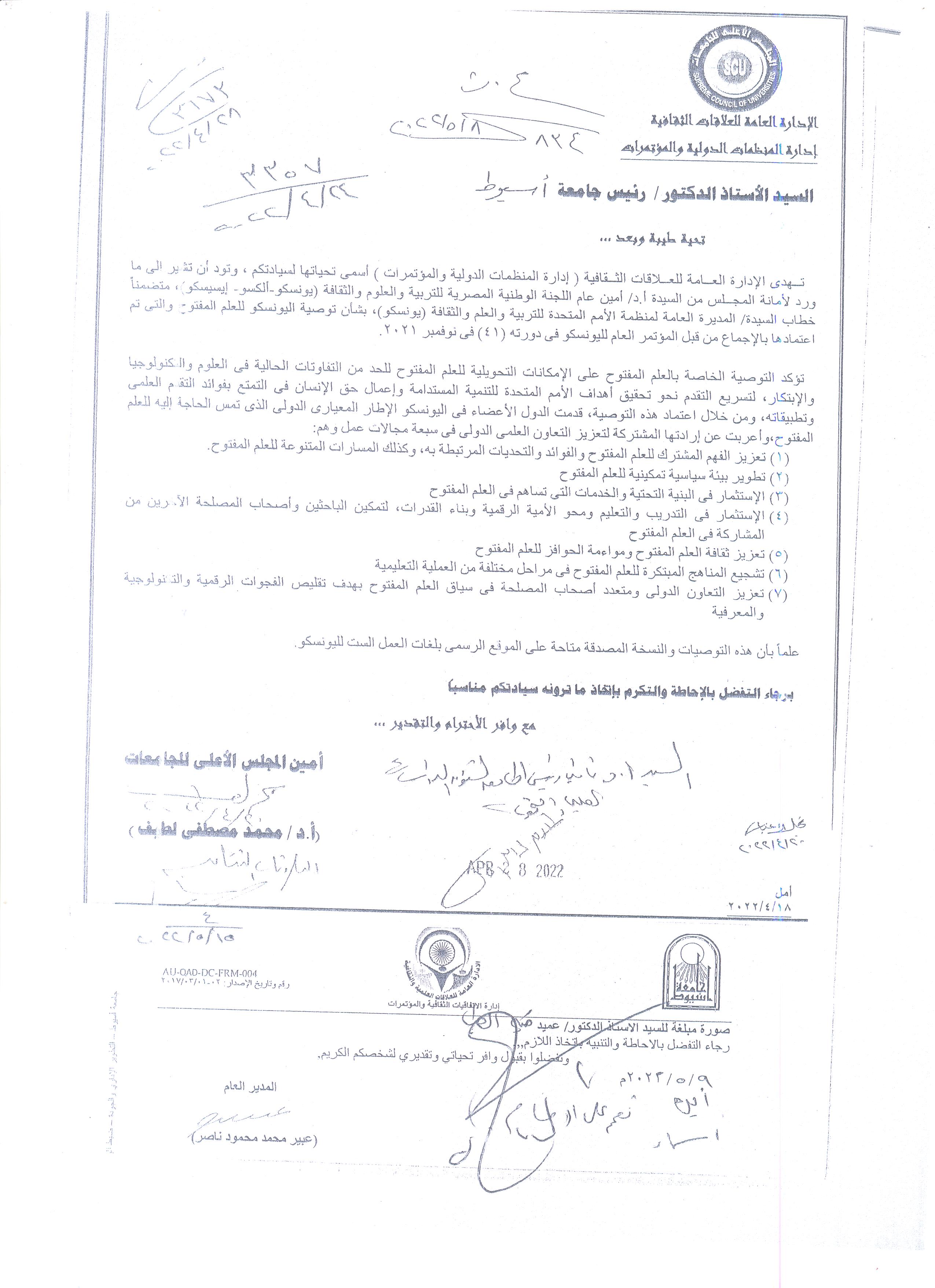
Declaration on the UNESCO Recommendation for Open Education at its 41st session in November 2021
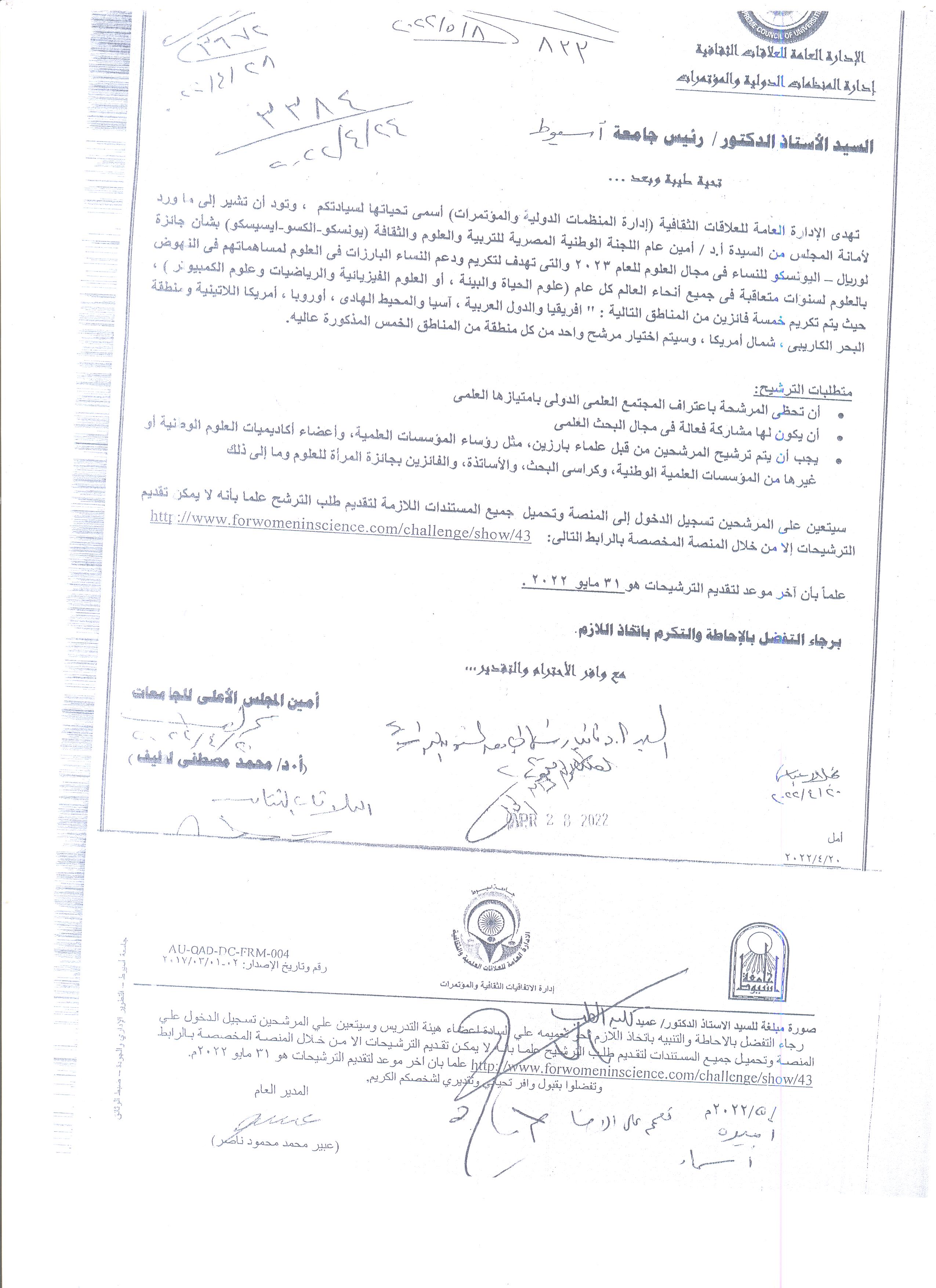
Announcement regarding the UNESCO L’Oréal Prize for Women in Science for the year 2023
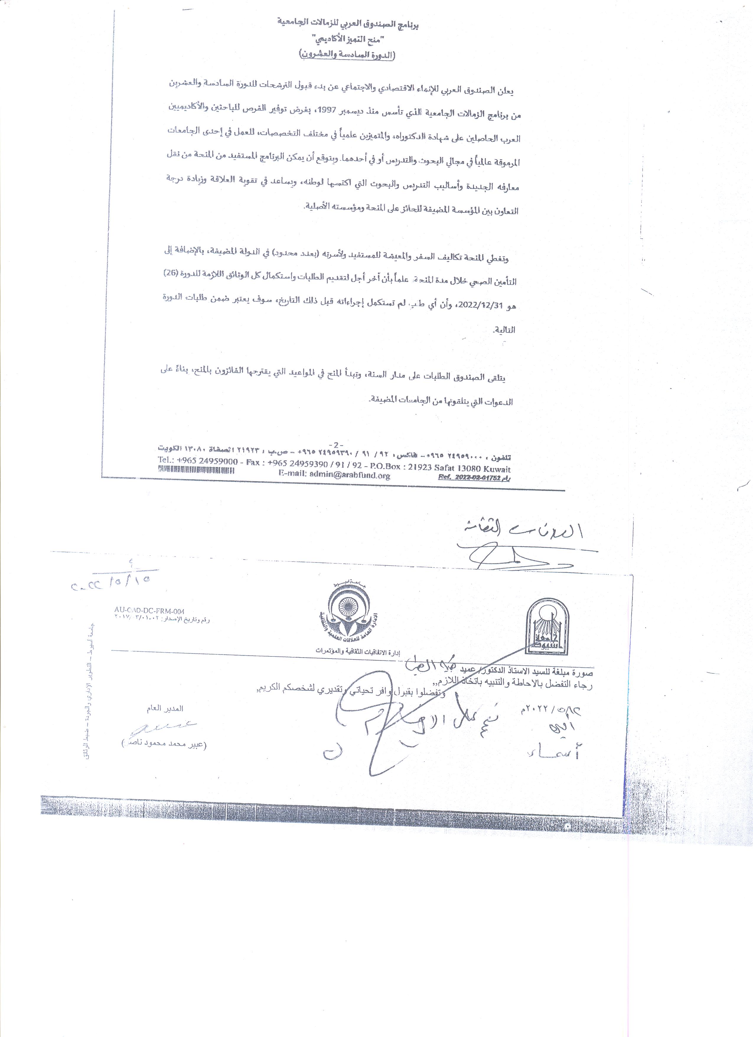
Announcement regarding the Arab Fund for University Fellowship Program (the twenty-sixth session)
Congratulations to the faculty members on the new promotions
Congratulations - Dr. Mohamed Ahmed Medhat - and - Dr. Ghada Kamal - Department of Tropical Medicine and Gastroenterology

أ. د/علاء عطية عميد كلية الطب ورئيس مجلس إدارة المستشفيات الجامعية.
أ. د/ اماني عمر وكيل الكلية لشئون التعليم والطلاب
أ. د/ يوسف صالح وكيل الكلية لشئون الدراسات العليا والبحوث
أ. د/ سعد زكي محمود وكيل الكلية لشئون خدمة المجتمع وتنمية البيئة.
يهنئون
د /محمد احمد مدحت
د /غادة كمال
وقسم طب المناطق الحارة والجهاز الهضمي
وذلك لدورهم ومساهمتهم في نشر أكبر دراسة مصرية مشتركة صادرة من ١٤ مركز ومستشفي جامعي بمصر عن خصائص أمراض القولون المناعية (القولون التقرحي ومرض كرونز) في مجلة Frontiers in Medicine (Q1)
و مبروك لكلية الطب وجامعة اسيوط
Congratulations - to the winning students of the Faculty of Medicine (the ideal student) at the university level
أ. د/علاء عطية عميد كلية الطب ورئيس مجلس إدارة المستشفيات الجامعية.
أ. د/ اماني عمر وكيل الكلية لشئون التعليم والطلاب
أ. د/ يوسف صالح وكيل الكلية لشئون الدراسات العليا والبحوث
أ. د/ سعد زكي محمود وكيل الكلية لشئون خدمة المجتمع وتنمية البيئة.
يهنئون الطلاب
الطالبة/ الشيماء محمود حسين الفرقة الثالثة
بفوزها بالطالب المثالي علي مستوي الجامعة
المركز الثاني
الطالبة/تسنيم محمد حسن الفرقة الثانية
بفوزها بالطالب المثالي علي مستوي الجامعة
المركز السادس
الطالب /مصطفي أحمد شاكر. الفرقة الثالثة
بفوزه بالطالب المثالي علي مستوي الجامعة
المركز السابع
ويعد هذا استمراراً لما يقدمه طلبة كلية الطب البشرى من إنجازات وبالتوفيق

Spastic Paraplegia 20 and Serine/Threonine Protein Kinase 31 Expression for the Detection of Colorectal Cancer
Research Abstract
Genetic alterations, including changes in the expression of spastic paraplegia 20 (SPG20) and serine/threonine protein kinase 31 (STK31), may play an important role in the carcinogenesis of colorectal cancer (CRC). Identification of such changes is suitable for the recognition of tumors at an early stage, which would significantly improve patient survival. While recent studies have identified that SPG20 and STK31 expression levels increase in CRC tissues, their use as a biomarker is yet to be investigated. Our aim was to determine whether circulating SPG20 and STK31 mRNAlevels could help distinguish between patients with CRC and healthy individuals. Additionally, we aimed to analyze the correlation between SPG20 and STK31 expression patterns and the tumor stage in patients with CRC.
Research Date
Research Department
Research Journal
Cell Physiol Biochem
Research Member
Research Pages
12
Research Publisher
Cell Physiol Biochem
Research Vol
56
Research Website
https://articles.cellphysiolbiochem.com/Articles/000509/PDF/000509.pdf
Research Year
2022
Is Prioritizing a Certain Method of Health Care Education of Patients Undergoing Percutaneous Suprapubic Catheterization Evidence-Based? A Systematic Review
Research Abstract
The quality of evidence of the superiority of a certain method of health care education of patients undergoing suprapubic catheterization is very low. The evidence-based research on this topic is not sufficient to develop comprehensive conclusions. Patient-centered, well-designed research is highly recommended.
Research Date
Research Department
Research Journal
Urologic Nursing
Research Member
Research Pages
79-92
Research Vol
42
Research Year
2022
The Authority of Gene Modifiers in β-Thalassemia Major and Its Relationship to the Pathophysiology of the Disease
Research Abstract
β-Thalassemia, an inherited red blood cell disorder, presents a significant health problem worldwide and is caused by defects in the β-globin gene, resulting in the reduction or absence of β-globin chain synthesis. That leads to blood transfusion dependency with its terrible complications. Polymorphisms at position-158 of XmnI-HBG2 on chromosome 11 and BCL11A site on chromosome 2p16 might be linked with elevated hemoglobin F (HbF) appearance, which may, in turn, improve β-thalassemia sternness. This study aims to walk around the amending effects of XmnI and BCL11A loci on HbF levels in Egyptian β-thalassemia patients. Material and Methods. A prospective case-control study of 70 multi-transfused β-thalassemia major patients and 22 controls was performed in the Paediatric hematology unit of Assiut university hospital from June 2019 till April 2021. PCR-RFLP was used to detect single nucleotide polymorphisms at XmnI and BCL11A site loci. Results. XmnI Polymorphism was detected in 9 of 70 patients and associated with higher mean HbF levels (53.48%) than patients without polymorphism (mean Hb level was 42.23%)(P-Value= 0.035). The frequency of CT heterozygous genotype was 8 (11.4%), TT homozygous genotype was (1.4%), while the wild genotype CC was detected in 61 (87.1%) of the cases. While BCL11A Polymorphism detected in 21 of 70 patients did not affect either Hb or HbF levels (P-Value= 0.26). The TT genotype frequency was 49 (70%), and TC heterozygous genotype was detected in 21 (30%) of patients. The CC genotype was absent. Conclusion: XmnI-158Gγ polymorphism, but not BCL11A …
Research Date
Research Department
Research Member