إنجاز آخر جديد لأبنائنا الطلاب فى مجال البحث العلمى وهو خير دليل على نجاح كلية الطب جامعة أسيوط في بناء جيل من الأطباء الباحثين القادرين على المنافسة عالميًا
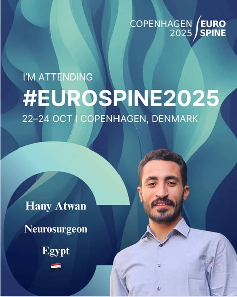
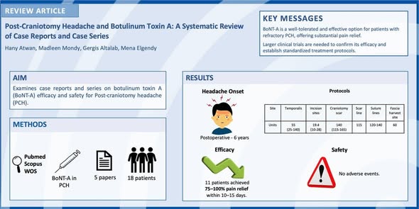
استطاع الطبيب المتدرب (طبيب امتياز) هاني سعيد موسى عطوان – دفعة 59 بكلية الطب جامعة أسيوط – تحقيق إنجاز بحثي لافت خلال شهر أكتوبر، بمشاركته في أربعة من أبرز المؤتمرات الطبية العالمية، وتقديمه 10 أبحاث علمية رائدة في تخصصات طب المخ والأعصاب، السكتة الدماغية، وجراحة العمود الفقري.
المؤتمرات التي شارك بها:
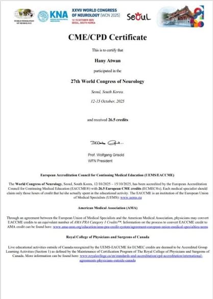
World Congress of Neurology – WCN 2025 (Seoul, South Korea)
أكبر تجمع عالمي لخبراء طب الأعصاب، ينظمه الاتحاد العالمي لطب الأعصاب WFN، ويضم مئات الجلسات العلمية التي تناقش أحدث الاكتشافات في مجالات السكتة الدماغية، والصرع، والأمراض العصبية.
EANS Annual Congress 2025 (Vienna, Austria)
الحدث السنوي الأبرز الذي تنظمه الجمعية الأوروبية لجراحي الأعصاب (EANS)، ويجمع نخبة من المتخصصين في جراحة الدماغ والعمود الفقري لعرض أحدث الأبحاث والتقنيات الجراحية المتقدمة.
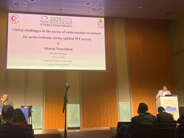
World Stroke Congress 2025 (Barcelona, Spain)
مؤتمر عالمي تنظمه المنظمة العالمية للسكتة الدماغية (WSO)، يُركز على التطورات الحديثة في الوقاية والعلاج وإعادة التأهيل لمرضى السكتة الدماغية بمشاركة واسعة من الباحثين والأطباء من مختلف دول العالم.
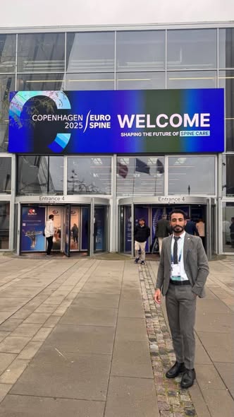
EUROSPINE 2025 (Copenhagen, Denmark)
أكبر مؤتمر أوروبي متخصص في أمراض وجراحة العمود الفقري، يجمع بين البحث العلمي والتطبيق العملي من خلال عروض تفاعلية وورش عمل حول أحدث أساليب العلاج وإعادة التأهيل.
وقد حصل الطبيب المتدرب على تكريم دولي مُشرّف خلال فعاليات مؤتمر EUROSPINE 2025، بـ جائزة EUROSPINE Early Career Award تقديرًا لتفوقه الأكاديمي ومشاركته المتميزة ضمن برنامج SPINE360 – Building Your Future in Spine Care، الداعم لفئة الباحثين الشباب وقادة المستقبل في طب وجراحة العمود الفقري.
تهنىء اسرة كلية الطب برئاسة الاستاذ الدكتور علاء عطية ، الطبيب المتدرب هانى سعيد بهذا الانجاز ، وتعبر عن اعتزازها وفخرها بابن كلية الطب جامعة أسيوط؛ لتشريفه الكلية في المحافل الدولية، والذي يعد وسام تقدير لكلية الطب؛ لما قدمه من جهد وأبحاث تخدم المنظومة البحثية في بداية حياته العملية.
 Do you have any questions?