Schedule for the academic year 2024/2025 - first semester - first level - eligibility
Schedule for the academic year 2024/2025 - first semester - first level - general
https://docs.google.com/spreadsheets/d/1vWc4s_V5LEsPrfpnGgqGCreJ-YNfq_mC/edit?usp=sharing&ouid=109287265863998872501&rtpof=true&sd=true
"The Objectives of the National Anti-Corruption Strategy"

The Faculty of Computers and Information at Assiut University hosts a seminar entitled "The Objectives of the National Anti-Corruption Strategy" to raise awareness of the risks of corruption and the need to combat it
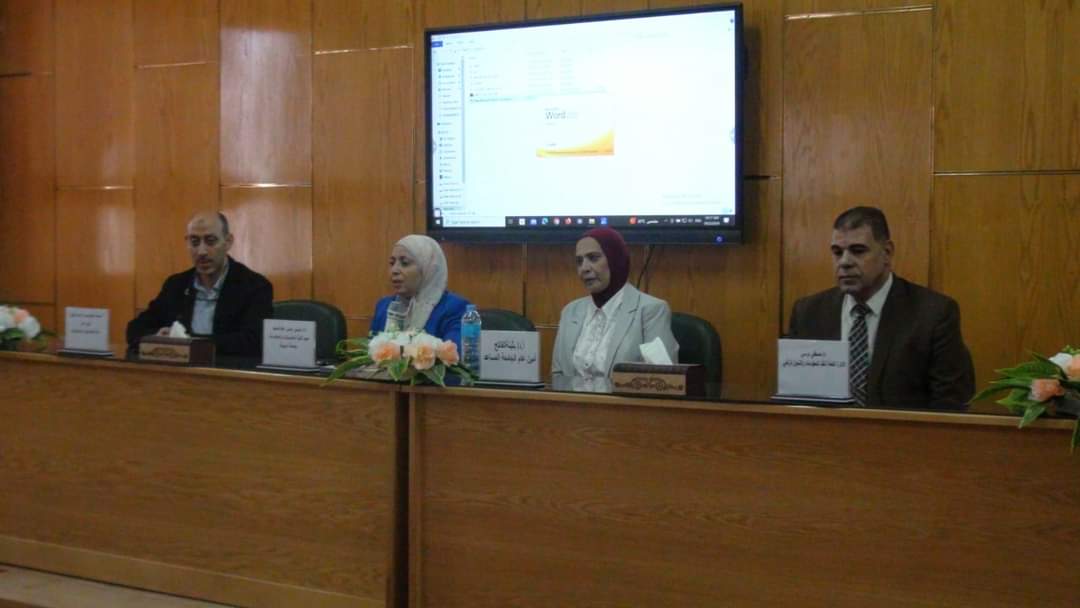
Under the supervision of Prof. Ahmed Al-Manshawi, President of Assiut University; The General Directorate of Information Systems and Digital Transformation, in cooperation with the General Directorate of skills Management and Development, organized an awareness seminar; as part of the series of seminars launched by Assiut University; to introduce the national anti-corruption strategy, on Sunday, September 22, at the Faculty of Computers and Information at the university.
Prof. Ahmed El-Minshawy, President of Assiut University, stressed the university's keenness to hold awareness seminars, which contribute to achieving national strategies aimed at combating corruption, and addressing all its forms, and its images, explaining that the symposium discusses the national anti-corruption strategy, in its third phase 2023-2030, and explains its main objectives, where the goal came; an efficient administrative apparatus that provides distinguished services to citizens; to enhance the efficiency and effectiveness of the administrative apparatus of the state; in line with the axis of transparency, and the efficiency of government institutions in Egypt's Vision 2030 for sustainable development on the one hand, and Egypt's plan for administrative reform on the other hand.
The symposium came under the supervision of: Mr. Shawkat Saber, Secretary General of the University, Dr. Butheina Al-Fatih, Assistant Secretary General of the University, Dr. Tayseer Hassan Abdulhamid, Dean of the Faculty of Computers and Information, Mr. Mohammed Abdul Mohsen, Secretary of the College, Dr. Mustafa Morsi, Director General of the General Directorate of Information Systems and Digital Transformation, and Coordinator of the Implementation of the Anti-Corruption Strategy for Assiut University.
Dr. Tayseer Hassan expressed her pleasure in hosting the symposium activities at the Faculty of Computers and Information, thanking all those responsible for the work of the Information Systems Department; for the efforts exerted in organizing events that are concerned with raising awareness, developing, and developing the academic staff and the administrative staff at the university.
Dr. Buthaina Al-Fatih explained: The university seeks to implement the national anti-corruption strategy, disseminate and establish the values and principles of transparency and integrity, at all levels, as well as developing and developing the skills of employees, in various sectors of the administrative apparatus; in order to create a positive work environment; contributes to achieving the university's goals and vision to develop work and improve administrative performance.
During the symposium, Dr. Mustafa Morsi discussed several axes, including: the definition of corruption, the United Nations Convention against Corruption, the initial anti-corruption strategy 2014/2018, the second anti-corruption strategy 2019/2022, the third anti-corruption strategy 2023/2030, the authorities concerned with anti-corruption, anti-corruption frameworks, including: the legislative, institutional and regulatory framework, and the regulatory framework for the authorities concerned with combating corruption, including: the Administrative Oversight Authority, the Central Accounting Authority, the Supervisory Authority for Technical Works, the Industrial Supervision Department, and the Pharmaceutical Regulatory and Research Authority.
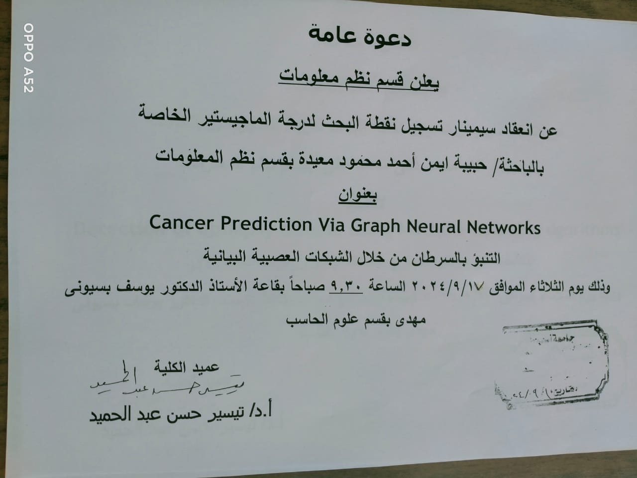
An invitation to hold a seminar to register the research point for the master’s degree for the researcher/ Habiba Ayman Ahmed Mahmoud, teaching assistant in the Information Systems Department.
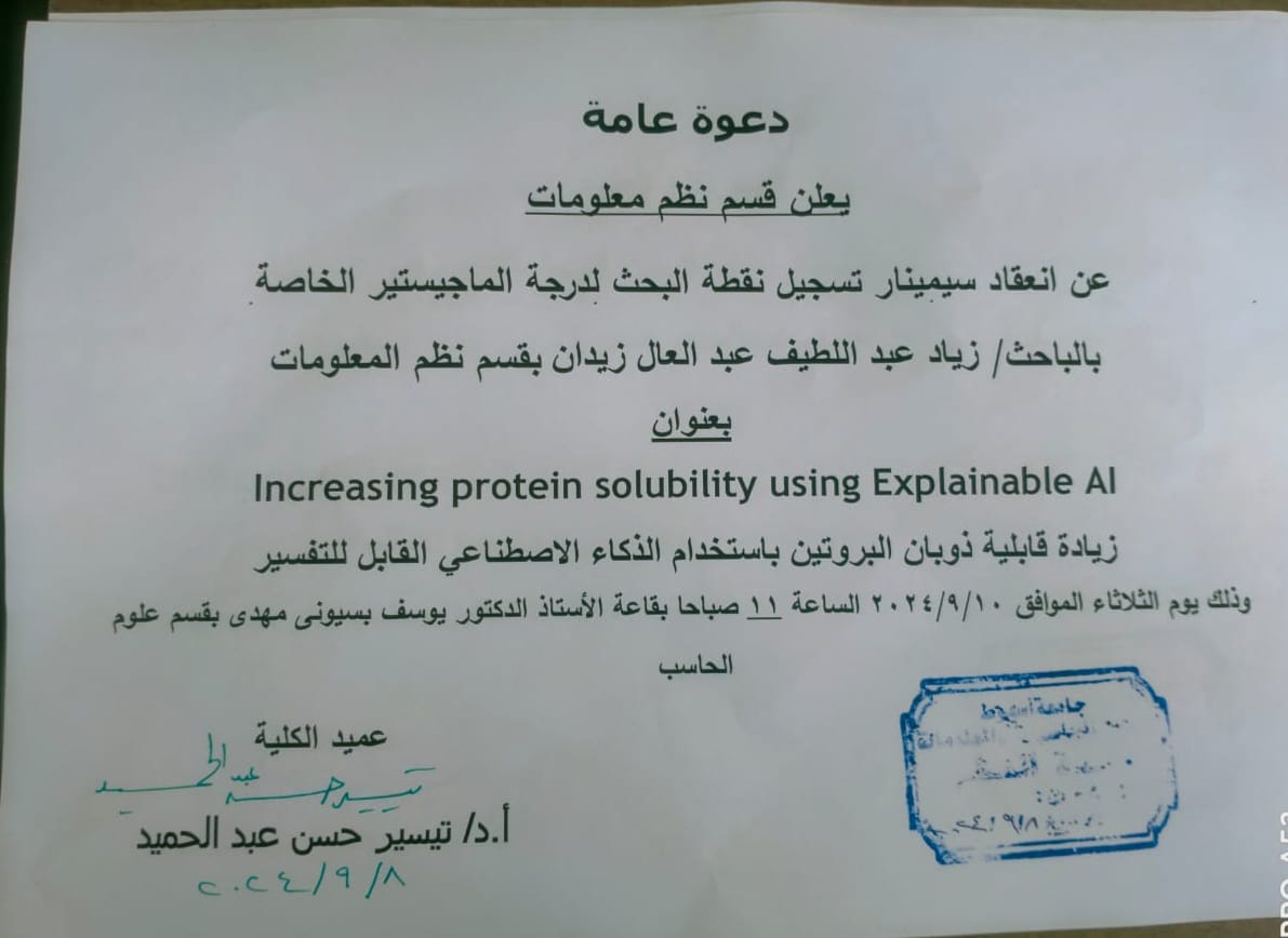
An invitation to register the research point for the master’s degree by researcher / Alaa Muhammad Zaki in the Information Systems Department
An invitation to register the research point for the master’s degree by researcher / Alaa Muhammad Zaki in the Information Systems Department

An invitation to register the research point for the master’s degree by researcher / Alaa Muhammad Zaki in the Information Systems Department

Important announcement for Assiut University students and graduates Info Session about Ejada Company Orientation
? طلاب وخريجى جامعة أسيوط
المهتمين بمجال ال IT ????
Attention Please
على هامش المائدة المستديرة الثالثة التى ينظمها المركز الجامعى للتطوير المهنى بجامعة أسيوط بالتعاون مع الجامعة الأمريكية بالقاهرة
UCCD Assiut University
بيقدملكم
يوم الخميس القادم ١٢ سبتمبر
الساعة ⌚١.٣٠ : ٢.٣٠م
بقاعة المناقشات رقم ١ المبنى الإدارى جامعة أسيوط
Info Session
مهمة جداً عن
Ejada Company Orientation
هتقدمهالنا
Eng. Haidy Hany
Manager, Campus Recruitment and HR Training Consultant
? خلال ال Session هيتم الإعلان عن ٥٠ فرصة وظيفية بشركة Ejada فى مجال تكنولوجيا المعلومات
?للتسجيل
https://docs.google.com/forms/d/e/1FAIpQLSd31-r5OvghMP2GKjirHGLIJxyfFzHf62c_Hc5-fGS26cZj0A/viewform?usp=sf_link
news category
Alumni