
Vaccination Campaigns for All Medical Crews in Assiut University
- وحدة علاج أعضاء هيئة التدريس للعاملين بالمستشفى الرئيسي و مستشفى ام القصور و مستشفى أسيوط الجديدة و مستشفى الطلبة

- وحدة مكافحة العدوى بمستشفى المسالك الدور السادس للعاملين بمستشفى المسالك

- وحدة مكافحة العدوى بمستشفى العصبية للعاملين بمستشفى العصبية

- وحدة التطعيمات بمستشفى الراجحي بالدور الأرضي للعاملين بمستشفى الراجحي

- وحدة مكافحة العدوى بمستشفى القلب الدور الأول للعاملين بمستشفى القلب

- وحدة العلاج بأجر بمستشفى الأسنان الدور الأول للعاملين بمستشفى الأسنان الجامعي

و جاري توفير التطعيم للعاملين بمستشفى الأطفال و مستشفى صحة المرأة و سيتم الإعلان عنها فور وصول الجرعات

كل الشكر للإدارة العليا و إدارات المستشفيات و إدارات التموين الطبي على الدعم و التعاون المستمر لتوفير التطعيم للاطقم الطبية و العاملين .

The Infection Control Unit in Assiut University Hospital launched vaccination campaigns for all medical crews against seasonal influenza. For more information about the places that offer this service, kindly read the provided list.
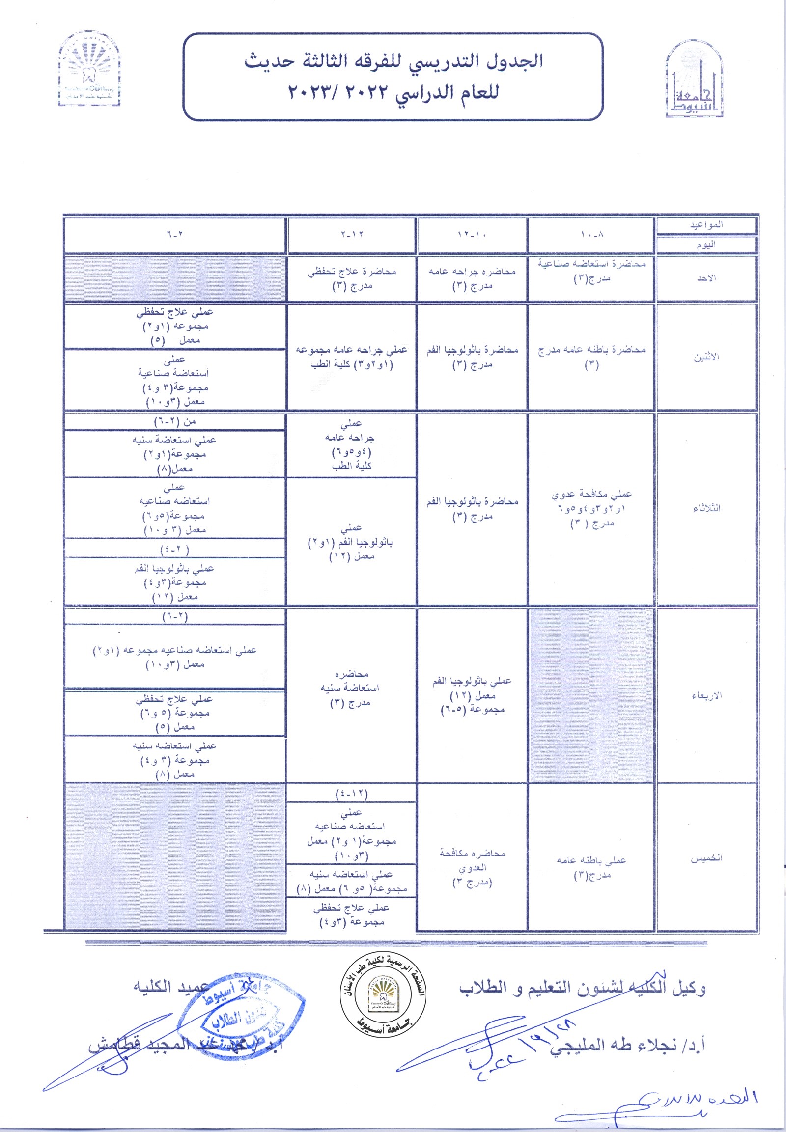
 

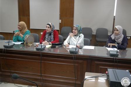
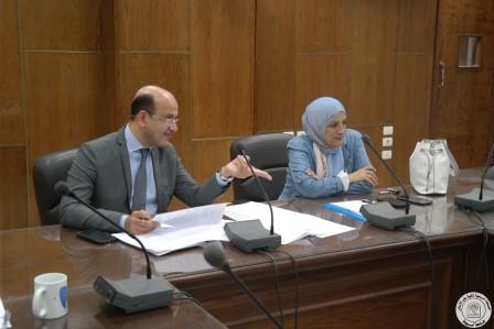
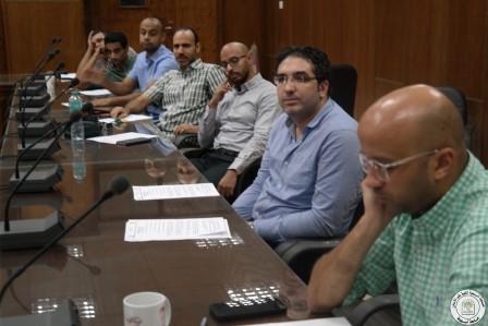
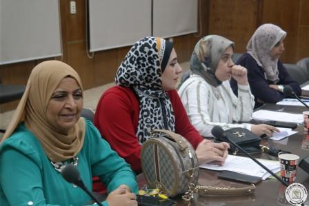
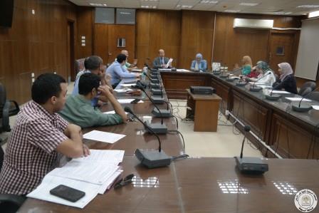
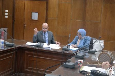
The Faculty Council held its 6th Meeting on Wednesday 29-9-2022

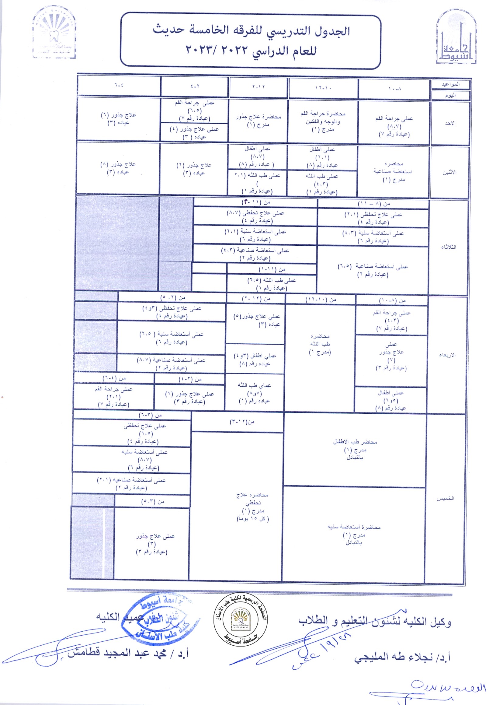
The Faculty Council held its 6th meeting on Wednesday 29-9-2022 to discuss the preparations made for the beginning of the new academic year 2022-2023 in the Faculties of Dentistry in Assiut University and in Assiut National University

.

.

.

.

.

.

EFFICACY OF R-ENDO AND E3-REROOT RETREATMENT ROTARY NITI SYSTEMS IN INTRACANAL FILLING MATERIAL REMOVAL

Research Abstract

ABSTRACT

Aim: To compare the effectiveness of R-Endo and E3-ReRoot retreatment rotary files in filling material removal and operating time.

Materials and Methods: Twenty extracted distobuccal roots of maxillary first molars were decoronated to a standard length of 12 mm. Root canal preparations were done using Fanta Blue (Fanta; ShangHai, China) till 0.04/25. Matching gutta percha cones of 0.04/25 were used with Adseal resin sealer (Meta Biomed, Chungcheongbuk-do, Republic of Korea) in warm vertical compaction technique. Teeth were left in an incubator at 37 °C and 100% humidity for 30 days. Cone beam computed tomographic images of all roots were obtained to confirm three-dimensional filling of the root canal space. Roots were divided into two groups according to the file used in retreatment; group A using R-Endo (Micro-Mega, Besancon, France) and group B using E3-ReRoot (IMD; ShangHai, China). Time required to remove the filling material was recorded in seconds. Teeth were then split into two halves using hammer and chisel and remaining filling material was calculated using stereomicroscope at 4X original magnification. Statistical analysis was performed using One-Way ANOVA and significance level was set at 0.05.

Results: E3-ReRoot was shown to be significantly more effective than R-Endo in intracanal filling removal. No significant difference was shown between both systems in the time needed to reach the working length.

Conclusion: E3-ReRoot was more effective in intracanal filling removal although both systems failed to totally remove gutta percha and sealer remnants from the root canals.

Research Authors
Tarek Medhat Elsewify* and H. M. Elhalaby**
Research Date
Research Department
Research File
د.هشام.pdf (699.62 KB)
Research Journal
EGYPTIAN DENTAL JOURNAL
Research Pages
3727-3733
Research Publisher
EGYPTIAN DENTAL JOURNAL
Research Rank
Local
Research Vol
65
Research Website
https://edj.journals.ekb.eg/article_76016.html
Research Year
2019

Evaluation of regenerative potential of dentin conditioning and naturally derived scaffold for necrotic immature permanent teeth in a dog model

Research Abstract

Summary

This study evaluated the outcome of partial exposure of dentin matrix to ethylenediaminetetraacetic acid (EDTA) and application of platelet-rich fibrin (PRF) scaffold on regeneration of necrotic immature permanent teeth in a dog model. The present study was carried out on 216 permanent immature roots in nine mongrel dogs aged 6-9 months. Pulp necrosis and periapical pathosis were induced in 180 roots. These roots were divided into five equal groups (36 roots each) according to the treatment protocol: group I: blood clot; group II: 17% EDTA solution and blood clot; group III: PRF; group IV: 17% EDTA solution and PRF; and group V: without treatment (positive control). The negative control group (group VI) represented 36 untouched normal roots for normal maturation. The groups were followed up for 1, 2 and 3 months (subgroups). Maturation of the roots was monitored by radiography and histopathology. All data were statistically analysed. Group IV exhibited the highest increase in root length and thickness, decrease in apical diameter, the highest score of vital tissue infiltration and least inflammatory scores. There was a significant difference regarding the increase in root length and thickness and decrease in apical diameter in all subgroups of the experimental and negative control groups (P ≤ .05). PRF has a better regenerative potential than the blood clot during treatment of immature permanent teeth with necrotic pulp. Inclusion of 17% EDTA solution as a final irrigation enhances the regenerative potential of both PRF and blood clot.

Research Authors
Hisham M. El Halaby1 | Ashraf M. Abu-Seida2 | Mervat I. Fawzy3 | Mona H. Farid4 | Hagar A. Bastawy3,5
Research Date
Research Department
Research File
د. هشام.pdf (1.75 MB)
Research Journal
International Journal of Experimental Pathology
Research Pages
264-276
Research Publisher
Wiley Online Library
Research Rank
International
Research Vol
101
Research Website
https://onlinelibrary.wiley.com/doi/10.1111/iep.12372
Research Year
2020

ROOT SURFACE STRAIN AND CRACKS DEVELOPMENT BY TWO RECIPROCATING NICKEL TITANIUM FILES

Research Abstract

ABSTRACT

Aim: The aim of this study was to determine root surface strain (RSS) developed by two reciprocating NiTi files and the resultant cracks.

Materials and Methods: Thirty mesial roots of mandibular molars were used. Ten roots were prepared using AF Blue R3 Reciprocating One-File System (FBR) (Fanta; ShangHai, China) for the mesiobuccal canal and WaveOne Gold primary (WOG) (Dentsply Tulsa Dental; Tulsa, OK, USA) for the mesiolingual canal. Ten roots were prepared using WOG and ten roots using FBR. An electrical strain gauge was fixed on the root surface connected to a strain amplifier. Maximum strain and time were recorded. Roots were cut transversely to evaluate for cracks.

Results: WOG has developed RSS of (84.16±52.57) and cracks (2/10); FBR has developed RSS of (128.33±128.55) and cracks (5/10) without statistically significant difference. Preparation time of WOG was (55.28±26.24) which was statistically significantly less than FBR (109±57.3).

Conclusion: Both files have developed RSS and cracks during canal shaping.

Research Authors
Tarek Medhat Elsewify* and H. M. Elhalaby**
Research Date
Research Department
Research File
د. هشام.pdf (503.91 KB)
Research Journal
EGYPTIAN DENTAL JOURNAL
Research Pages
2689:2694
Research Publisher
EGYPTIAN DENTAL JOURNAL
Research Rank
Local
Research Vol
66
Research Website
https://edj.journals.ekb.eg/article_120885.html
Research Year
2020
Subscribe to