

Section Groups for Fifth Year Students 2022-2023
https://drive.google.com/file/d/1IT7UzwuJodbOWi1jEGAHc6BfdPH-hbPm/view?usp=sharing
Section groups for fifth year students 2022-2023. For more information, kindly visit the provided link.
https://drive.google.com/file/d/1IT7UzwuJodbOWi1jEGAHc6BfdPH-hbPm/view?usp=sharing
Section groups for fifth year students 2022-2023. For more information, kindly visit the provided link.
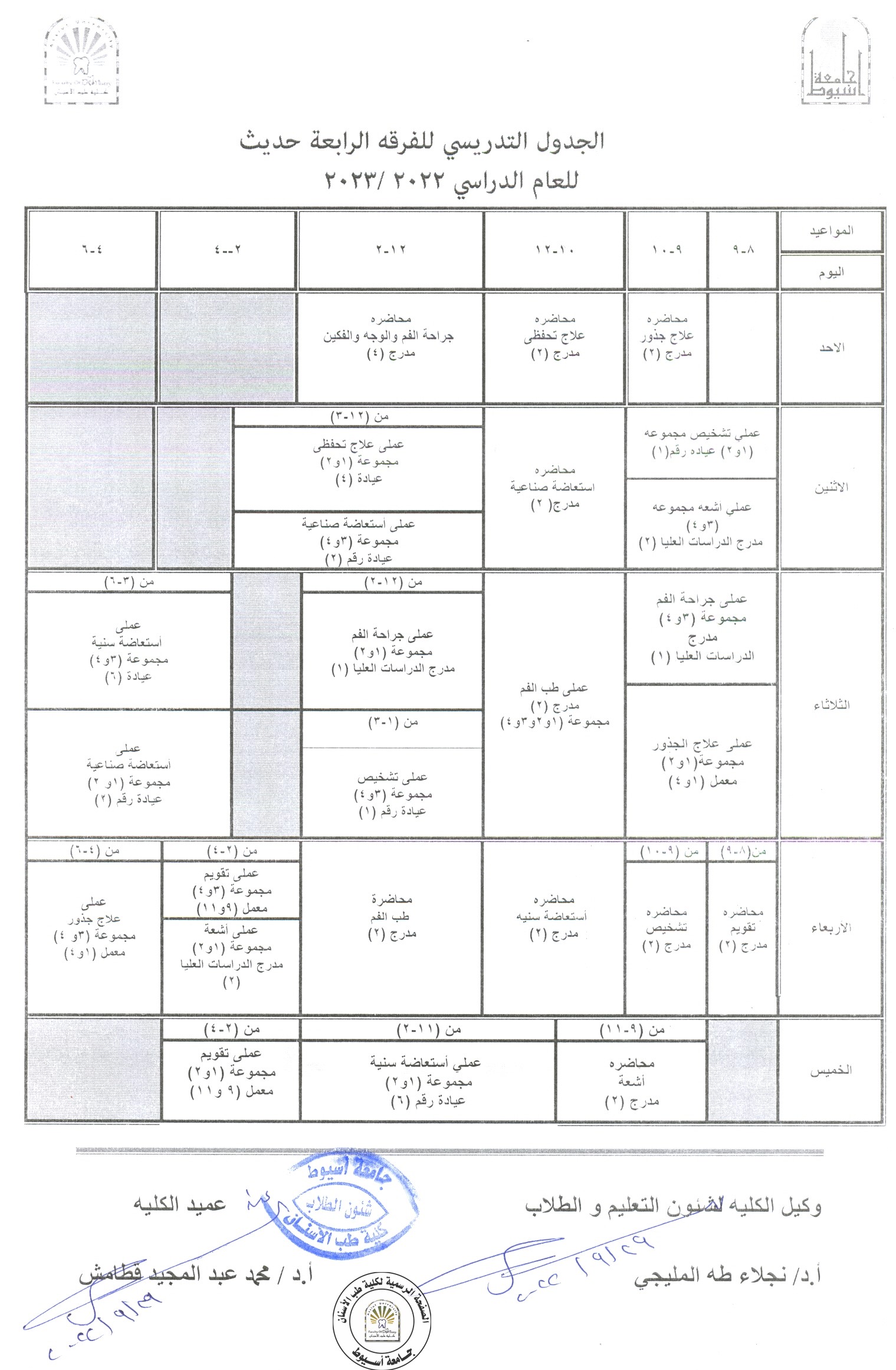
The New Schedule for Fourth year students (new system) 2022-2023.
Background
Diabetes mellitus is a generalized notorious chronic disease characterized by hyperglycemia. Salivary glands play an important role in the oral health maintenance by production of saliva. One of the most common diseases that compromise salivary gland function is diabetes mellitus leading to xerostomia.
Aim
The aim of this study was to determine the histological findings and immunohistochemical expression of cytokeratin 17 (CK17) in normal and diabetic submandibular gland to give an explication about the pathological effects of diabetes mellitus on the intracellular structures of the gland parenchyma.
Materials and methods
Twelve healthy male albino rats were utilized in the experiment and divided into two equal groups (normal and diabetic). Forty‑five days postinduction of diabetes, submandibular gland was dissected out and prepared for both histological and immunohistochemical studies.
Results
Histological results confirmed that, submandibular gland of diabetic rats undergoes rapid, progressive and severe atrophy of the parenchymal elements accompanied by increases in the fibrous component and presence of fatty degeneration. The atrophied gland was characterized by loss of the gland architecture with the presence of degenerated acini, dilated duct system and formation of duct-like structure. Expression of CK17 using immunoperoxidase technique revealed a mild to moderate cytoplasmic expression in all duct cells and some acinar cells as well as the granular convoluted tubules of normal gland either diffuses or concentrated lateral and basal to the nucleus compared to strong expression of diabetic glands that concentrated at apical part of cells.
Conclusion
The intensity and diffusion of CK17 expression in our results foretells the pathological influence of diabetes mellitus to the intracellular filaments of salivary gland parenchyma that interfered with production and/or secretion of saliva leading to xerostomia.
Abstract
Background: Increasing the prevalence of oral Candidal growth is associated with the habit of smoking and increased with denture wearing, and many of these carry a potential risk for the development of denture-related stomatitis. Shisha smoking in the rural areas in Egypt is more prevalent than cigarettes, especially in old males. There have been no studies that report the prevalence of candidal growth and denture related stomatitis under complete dentures in shisha smokers, in rural regions, of south Egypt.
Materials and Methods: A total of 30 edentulous participants were selected and examined to assess any oral mucosal changes (15 were shisha smokers and 15 were non-shisha users). complete dentures were constructed, For every patient, oral examination and samples were collected at the following intervals: before denture insertion, one, three, and six months after denture adaptation.
Results: Shisha smoking affect significantly the mean count at different time interval (1,3,and 6 months) while the non shisha users was observed to show significant increase later at 3 and 6 months. Confluent growth was not observed among non- shisha users while it appears in about 7% of the shisha smokers group. Moderate and severe inflammatory grades of palatal mucosa was noticed among shisha smokers compared to mild grade only seen in one case of non-shisha users after 6 months of insertion.
Conclusion: shisha smoking increases the prevalence of candidal growth under complete dentures and increases the probability of denture-related stomatitis to occur.
ABSTRACT
Midkine is a heparin-binding growth factor that promotes the proliferation, survival, migration and differentiation of various target cells. Midkine plays an important role in tumorigenesis and tumor progression, and is overexpressed in many human malignant tumors. The aims of this study were to evaluate the expression of von Willebrand Factor (vWF) and Midkine (MK) factor in oral squamous cell carcinoma (OSCC).And to correlate the expression of both (vWF) and (MK) with some clinicopathological data and lymph node metastasis. Immunohistochemical showed that MK protein expression was significantly higher in specimens of carcinomas with lymph node metastasis. Furthermore, vWF expression tended to be higher in cases that exhibited high expression of MK. These results suggest that MK may play important role in tumor’s progression and angiogenesis.
ABSTRACT
Objectives: Cell structure changes due to oxidative stress, and the response of cells to this change depends on the amount of oxidative stress. Generally, severe oxidative stress causes cell death. This study evaluates the oxidative therapy of curcumin on Hep-2 cell line.
Materials and Methods: Laryngeal squamous cell carcinoma cell line (Hep-2) cells were treated with curcumin for 48 h. Then, cell viability was tested using MTT assay, and the amount of Cox-2 and ROS (reactive oxygen species) was measured by enzyme-linked immunosorbent assay (ELISA) assay; the amount of MDA was measured by colorimetric/fluorometric assay. Finally, slides were prepared for hematoxylin and eosin (H & E) staining for microscopy.
Results: The data revealed that curcumin had a cytotoxic effect on Hep-2-treated cells. Furthermore, curcumin downregulates Cox-2 and increases the accumulation of intracellular ROS and MDA levels compared with control untreated cells. Additionally, Hep-2-treated cells showed apoptosis.
Conclusions: Our findings suggest that curcumin can be an oxidative therapy for laryngeal squamous cell carcinoma and induce apoptosis. Also, curcumin has anti-inflammatory activity in Hep-2 cells as it can decrease Cox-2 levels in cells. Thus, curcumin is a potential antiproliferative and therapeutic agent.
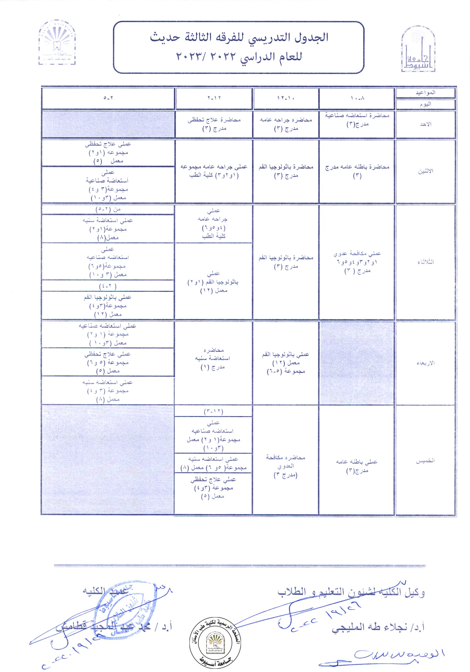
The New Schedule for Third Year Students (New System) 2022-2023.
من خلال الرابط التالي :
https://drive.google.com/file/d/1DJ0qZtaDYgTr5CJrq7M9_dxnAEuOeQkj/view?usp=sharing
Section Groups for First Year Students 2022-2023.
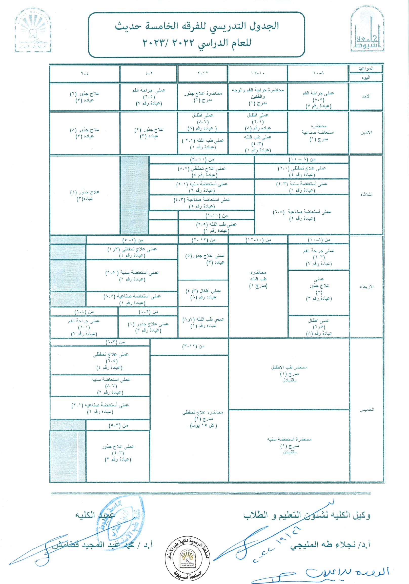
The New Schedule for Fifth Year Students 2022-2023.
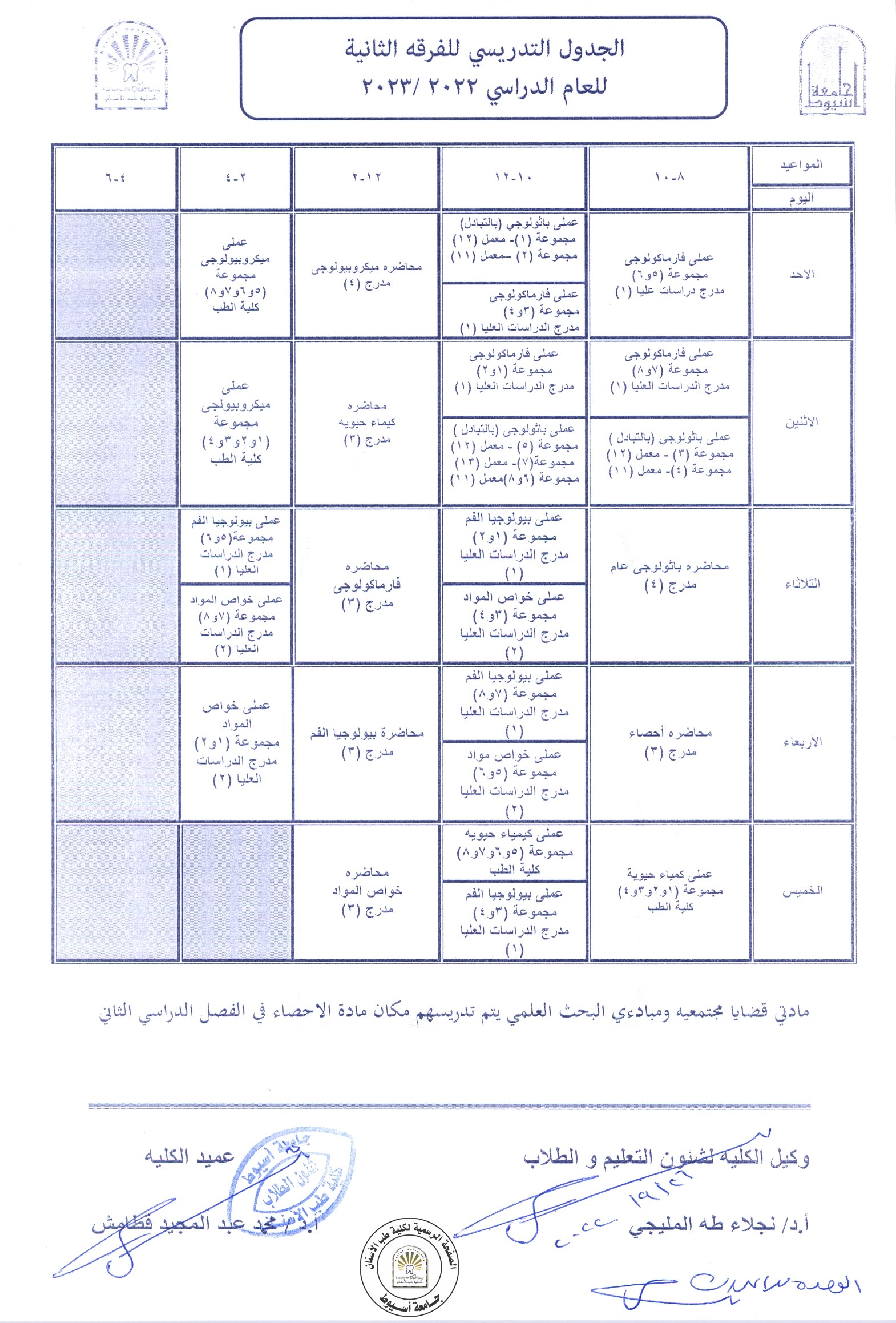
The Latest Schedule for Second Year Students 2022-2023.