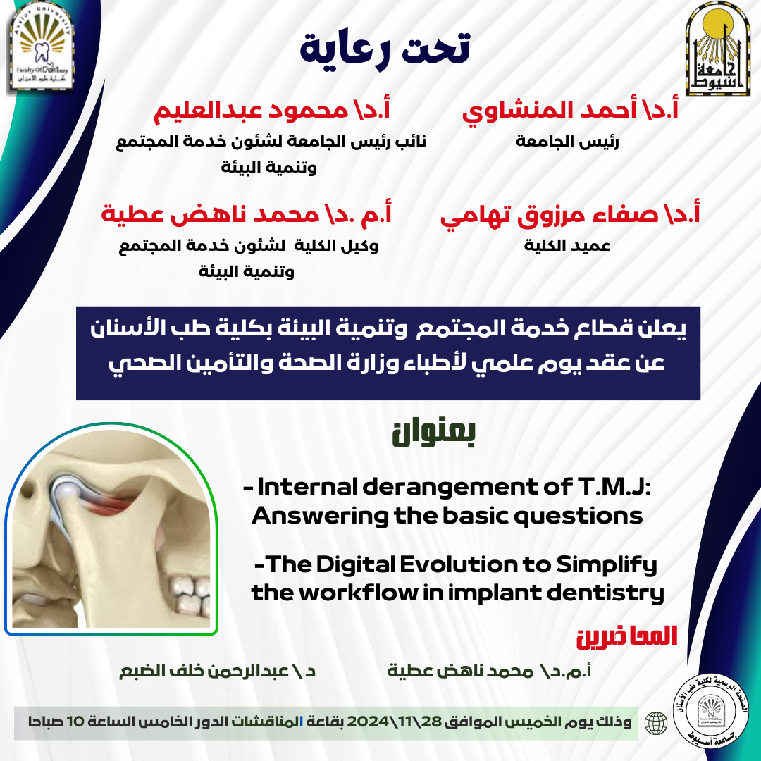
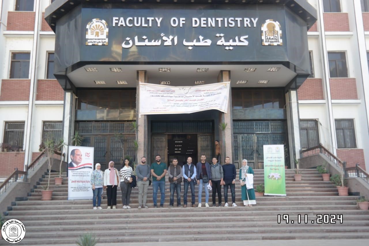
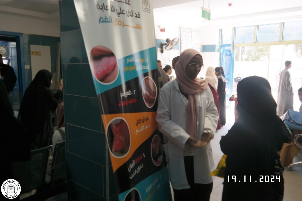
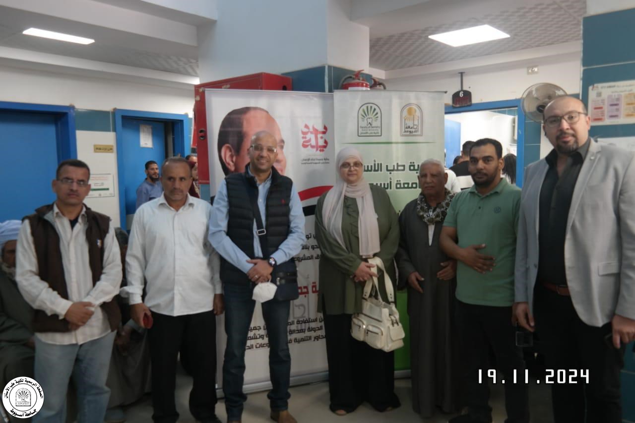
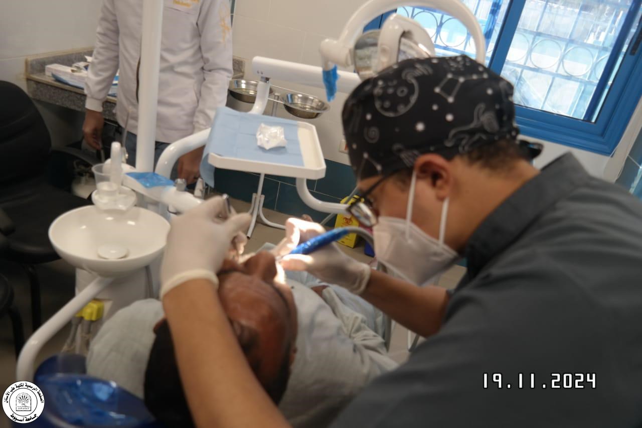
Dental Interns to Meet Head of Dental Hospital

The dental interns (2024-2025) are invited to attend a meeting with the Head of the Dental Hospital on Sunday, October 28, 2024, at 9:00 a.m. in the Postgraduate Studies Hall.

The dental interns (2024-2025) are invited to attend a meeting with the Head of the Dental Hospital on Sunday, October 28, 2024, at 9:00 a.m. in the Postgraduate Studies Hall.
| Abstract | |
| Introduction: The most prevalent malignant tumor of the head and neck region is squamous cell carcinoma. The bad prognosis and low survival rate are mainly related to oral cancer patients. Developing chemopreventive techniques has been crucial to reduce the incidence of oral cancer. Chemoprevention with natural products has emerged as viable preventive options. Salvia rosmarinus and Coriandrum sativum have been studied and discovered to have powerful anticancer properties. Aim of the study: The present trial was proceeded to study the effects of salvia rosmarinus and coriandrum sativum during experimental hamster buccal pouch carcinoma induction; using PCNA as cancer proliferative cell marker. Material and Methods: A total of 60 adult male hamsters were divided into 5 equal groups. Group I, biopsies of normal pouch mucosa were obtained as a negative control. All remaining groups were painted by DMBA as a carcinogenic agent. Group II, as a positive control group, was only painted by DMBA. Group III received topically salvia rosmarinus oil. Group IV received topically coriandrum sativum oil. Group V received both oils together. After 16, 24, and 32 weeks, 2 to 4 animals from every group had been sacrificed. Tissue lesions were prepared, and paraffin sections were stained with primary antibody for PCNA immunohistochemical evaluation. Results: The salvia rosmarinus and coriandrum sativum; either alone or in combination, significantly reduced the carcinogenic effect for DMBA-induced oral carcinogenesis. Conclusion: Salvia rosmarinus and coriandrum sativum have a chemopreventive considerable action versus oral cancer development, and their synergetic action is considerably more effective. |
ABSTRACT
Purpose: This study was performed to introduce, evaluate, and compare various novel assessment protocols designed for straightforward, reliable, and reproducible measurement of alveolar bone levels. These protocols are intended for standardized periodontal assessment and follow-up, utilizing cone-beam computed tomography (CBCT) images and manipulation of Digital Imaging and Communications in Medicine (DICOM) viewer software.
Materials and Methods: Two experienced oral and maxillofacial radiologists developed 5 distinct radiographic measurement protocols. These techniques were established to assess the alveolar bone level of a periodontally affected upper central incisor using a method that is consistently repeatable across observers. Two additional assessors, blinded to the details of the study, independently applied the protocols to retrieved DICOM files that met the eligibility criteria. A scoring system with 3 subscores was created and used to compare the protocols.
Results: Statistically excellent inter-observer reliability was observed for all protocols, other than protocol 1, which demonstrated moderate reliability. The average discrepancy between measurements taken by the 2 observers was 1.2 mm for protocol 1, 0.81 mm for protocol 2, and less than 0.5 mm for the remaining 3 protocols. All approaches except protocol 4 were straightforward to apply.
Conclusion: This study introduces multiple reliable protocols for the evaluation of periodontal bone levels that ensure consistency across observers. Based on the findings, the double axial lines and incisocrestal distance protocols are recommended. These new assessment approaches, along with any future modifications, may be useful in periodontal assessment, dental implant follow-up, orthodontic evaluation, research, and artificial intelligence model generation. (Imaging Sci Dent 20240073)
Abstract:
Background: This research was designed to assess the nasopalatine canal shape, size as well as anatomic variations according to age & gender in a sample of the Egyptian population utilising CBCT .
Material and Methods: This research was conducted on 100 CBCT scans belonging to 31 males & 69 women, with mean age of (35.8 ± 12.3) years. Inclusion and exclusion criteria as well as ethical issues were designed. CBCT scans were classified according to gender (male and female) and according to age (18-40 year and 41-61 year). All nasopalatine canals were evaluated to assess nasopalatine canal shape in coronal view and sagittal view as well as measurements of the nasopalatine canal in the sagittal & axial cross-section CBCT. The data were collected, computed, tabulated and statistically analyzed.
Results: Was observed no statistically substantial association amongst gender & NPC shape. Also, was noted no statistically substantial association amongst age group and NPC shape. Males showed statistically significantly higher mean diameter of incisive foramen and nasopalatine canal length than females. Subjects aged 18 – 40 years old showed statistically significantly lower mean antero-posterior diameter of nasal foramen, medio-lateral diameter of incisive foramen as well as canal diameter than those aged 41 – 61 years old.
Conclusions: The most frequent NPC shape in coronal view is variation of the Y-type of NPC in half of participants followed by single canal and the least was 2 parallel canals. The most frequent NPC shape in sagittal view is funnel shape (29%) followed by cylindrical shape (28%). The antero-posterior diameter of the incisive foramen & the nasopalatine canal length are higher in men than women. The antero-posterior diameter of the nasal foramen, the medio-lateral diameter of the IF and the NPC increase with aging.
ABSTRACT
Aim: The aim of this study was to evaluate the immediate implant placement versus delayed implant placement 3 months later of the splitting of thin alveolar bone sites using flapless crest splitting technique.
Subjects and Methods: This study was designed as a prospective clinical trial of 35 implants over 8 patients with 16 alveolar splitting sites. Eight sites were splitted and immediately implanted and submitted to group I, the another eight sites were splitted and implanted after 3 months they were submitted to group II, After alveolar ridge spiltting both groups were grafted via xenografts and covered with collagen membranes for guided bonr regeneration (GTR). For both groups we clinically assessed insertion torque, Implant stability quotient (ISQ) and radiographically horizontal bone gain and bone density at 3 intervals postoperatively, 3 and 6 months.
Results: The mean ISQ and insertion torque ± SD in group II was higher than group I. Group I showed a higher increase in bone gain than group II.
Conclusion: Immediate implantation after piezotome ridge splitting can be a useful procedure in ridges which have low bone quality and a thin cortex. Delayed implantation after piezotome ridge splitting is recommended when the initial stability of the implants is predicted to be poor. Both techniques using piezotome alveolar ridge splitting are effective in avoiding adjacent nerve injury.
ABSTRACT
Aim: This study was conducted to compare Computed guided arthrocentesis with conventional arthrocentesis followed by I-PRF prolotherapy in the treatment of TMJ internal derangement.
Subjects and methods: the study was carried out on 20 patients divided into two groups: Group I, patients underwent arthrocentesis followed by I-PRF injection using conventional TMJ injection technique with the aid of facial anatomical landmarks. Group II, patients underwent arthrocentesis followed by I-PRF injection using a CT-guided 3D printed surgical guide. Pain scores measured and MIO, preoperatively, immediately after the procedures, 1 week, 1 month, and 3 months. MRI was made 1 week and 3 months after the procedures. The number of attempts of needle insertions of 1st & 2nd needle (Right -Left) were assessed and the Time of operation and pain during the operation were assessed. Results: a significant decrease in pain during procedures, immediately post-operative, 1 week and 1 month post-operative, number of insertions and relocation of the needles in each joint, and time of the procedures in the guided arthrocentesis group compared to the conventional arthrocentesis group. On the other hand, there was no significant difference between both groups in MIO, preoperative pain after 3 months.
Conclusion: Digital arthrocentesis and prolotherapy of TMJ showed superiority and could replace the current conventional and prolotherapy types. Intra-articular PRF injection after arthrocentesis is an effective treatment method for the pain relief of TMJ internal derangements.
ABSTRACT
Aim: Our investigation aimed to evaluate the prevalence and patterns of impacted third molars
in a sample of Upper Egyptian patients and explore their association with different sagittal skeletal
relationships.
Materials and methods: A total of 352 pre-treatment panoramic radiographs (OPGs) and lateral
cephalograms were retrospectively reviewed. The patients, aged 18 to 40 years, were classified into
three skeletal groups based on the ANB angle. The number and angular position of impacted third
molars were assessed. We used Winter’s classification to find out impacted third molars angulation.
Results: Compared to Class I and Class III, the results disclosed a higher prevalence of third
molar impaction in Class II malocclusions. Additionally, Class II individuals exhibited a higher
prevalence of mesioangular and distoangular impactions, while Class III patients showed a greater
tendency for horizontal impactions in the mandible. Vertical impactions were more common in
Class II individuals. Furthermore, gender differences were observed, with females exhibiting
a higher percentage of impacted molars across all skeletal classes.
Conclusion: Class II malocclusions had higher mesioangular and distoangular impaction rates,
while Class III malocclusions were prone to mandibular horizontal impactions. Gender-related
differences showed females had a higher incidence of impacted molars across skeletal classes.
Understanding the link between skeletal class and third molar impactions is vital for improved
orthodontic planning.
ABSTRACT
Objective: To compare with the aid of cone beam CT the effect of treatment with clear aligner versus fixed orthodontic appliances on the external root resorption in upper teeth.
Patients and Methods: Fourteen individuals, aged between 15 and 25, took part in the study. Research randomization was used to assign patients who satisfied the inclusion criteria at random to the aligner and fixed groups.
Results: There were statistical significant differences in root volume and linear measurements of both groups (Paired t-tests were used between T0 and T1). Independent t-tests were used to compare the mean changes of root volumes and linear measurements between the two groups).
Conclusion: There is a significant root resorption in both clear aligner and fixed orthodontic appliance groups. The difference between both groups was statistically insignificant.

The Scientific Committee at the Faculty of Commerce announces a scientific competition titled "Cinema of Science."
Competition Guidelines:
Competition Details:
Prizes:
Registration:
Interested participants should register with the faculty's scientific activity coordinator.

The Scouting and Public Service Unit at the Central Youth Welfare Administration announces the organization of the 43rd Scouting and 29th Guiding Courses for the university's scouts and guides.
Dates: Thursday, November 21, 2024, to Saturday, December 7, 2024
Location: The university's permanent scouting camp
Interested students should register with the coordinator of the Scouting and Public Service Unit at the Faculty of Dentistry's Youth Welfare Administration.