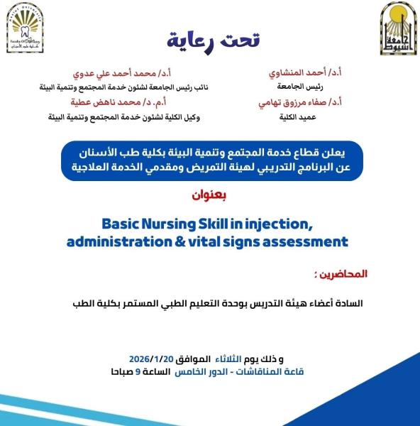
تحت رعاية
أ.د أحمد المنشاوي - رئيس الجامعه
أ.د محمد عدوي - نائب رئيس الجامعه
لشئون خدمة المجتمع و تنمية البيئة
أ.د صفاء تهامي - عميد الكلية
أ.د محمد ناهض عطيه - وكيل الكلية لشئون خدمة المجتمع و تنمية البيئة
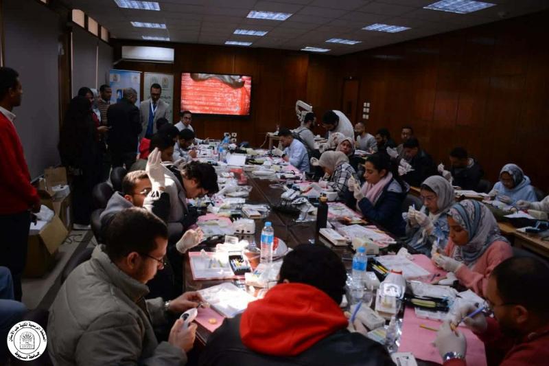
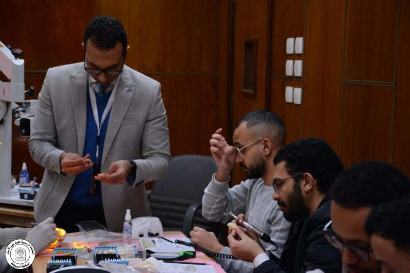
بنجاح عظيم تم بحمد الله و فضله الإنتهاء من ثالث ورش العمل التي نظمتها وحدة التعليم الطبي المستمر بالكلية بالتعاون مع قسم العلاج التحفظي علي مدار يومين ( ١٢ و ١٣ يناير ) في أساسيات الحشوات التجميلية للأسنان الأمامية تحت عنوان
(Resin Composite : The State of Art)
، تأتي هذه الورشة ضمن خطة قطاع خدمة المجتمع و تنمية البيئة لعقد سلسلة من ورش العمل علي مدار العام الجامعي ٢٠٢٥ - ٢٠٢٦ في إطار دور الكلية المجتمعي لرفع كفاءة أطباء الأسنان في صعيد مصر و المنتسبين إلي الكلية
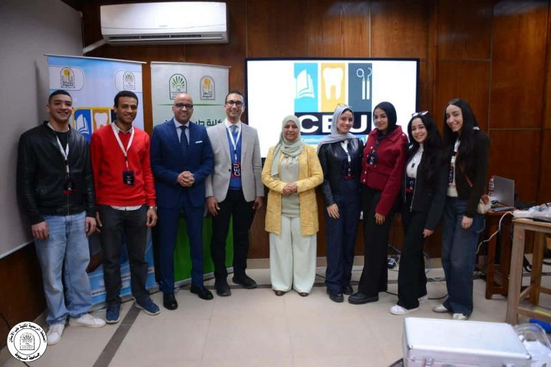
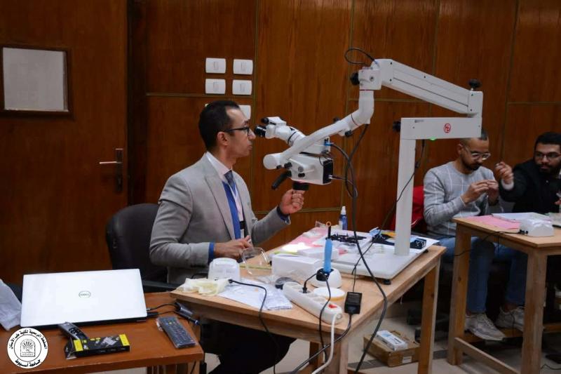
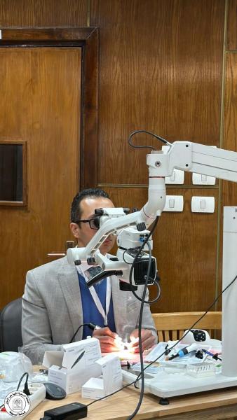
حاضر في ورشة العمل د. محمود القاضي - مدرس العلاج التحفظي و مدير وحدة التعليم الطبي المستمر - كلية طب الأسنان جامعة أسيوط و
بمشاركة متميزة من أعضاء الهيئة المعاونه بقسم العلاج التحفظي بالكلية
شارك في ورشة العمل أكثر من ٣٠ طبيب حيث تضمنت محاضرات نظريه و ورشة عمل عن أحدث الأساليب للتعويض التجميلي للأسنان الأمامية
خالص الشكر و التقدير و العرفان
د. محمود القاضي - مدير وحدة التعليم الطبي للمجهود العظيم و إسهاماته المشكورة محاضرا علي مدار اليومين و مشرفا علي ورشة العمل
خالص الشكر و التقدير و العرفان
- أعضاء الهيئة المعاونه بقسم العلاج التحفظي
- الشركات الراعيه لرعايتها المتميزه و إسهاماتها في خروج ورشة العمل بالشكل الأمثل
3M-Summit Store
Dental Capital
EIO - Ivoclar Distributor - Egypt (authorized distributor Eio)
الجزء الثاني من ورشة العمل سينعقد يومي الاثنين و الثلاثاء ٩ و ١٠ فبراير
للحجز و الاستعلام … التواصل مع وحدة التعليم الطبي المستمر علي الرقم
01030546900