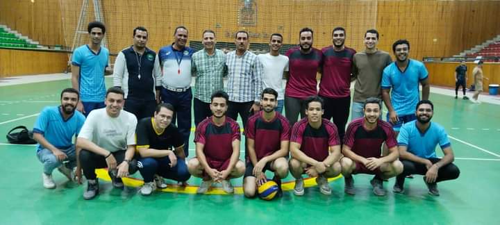
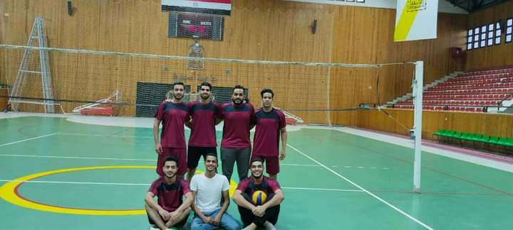
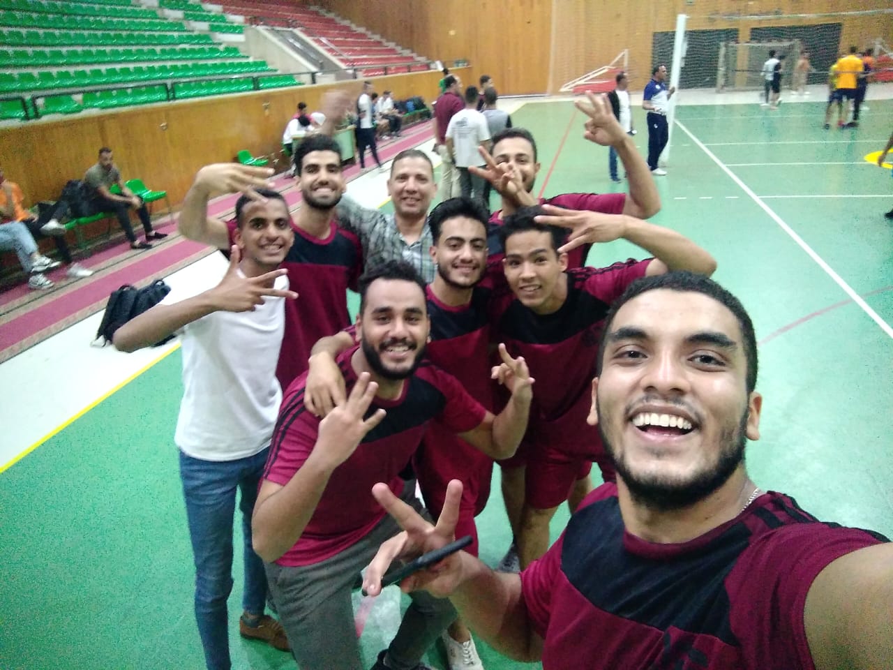
Congratulations - The Faculty volleyball team won third place at the Assiut University level
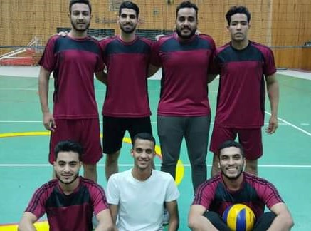
تحت رعاية السيدة الأستاذ الدكتور / مديحه درويش عميد الكلية والمشرف العام علي الانشطه الطلابيه بالجامعة
والسيد الاستاذ الدكتور / مؤمن عبد العظيم وكيل الكلية لشئون التعليم والطلاب
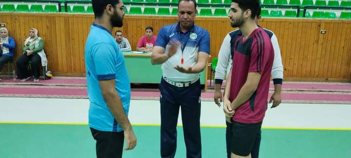
فوز فريق الكلية للكرة الطائرة بالمركز الثالث علي مستوي جامعة أسيوط بعد فوزه علي كلية الصيدليه
مشرف الفرق ا. محمد حسن
news category
student