Skip to main content
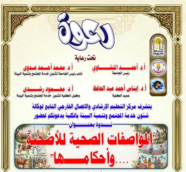
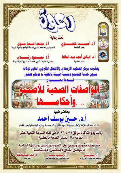
Invitation: Seminar entitled "Health Specifications and Rulings for Sacrificial Animals"
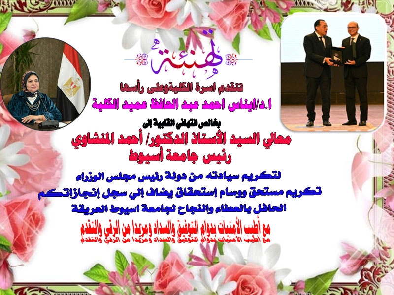
Congratulations from the Dean of the College to His Excellency the University President on the Prime Minister's honoring of him.
T cell subsets, regulatory T, regulatory B cells and proinflammatory cytokine profile in Schistosoma haematobium associated bladder cancer: First report from Upper Egypt
Molecular characteristics and zoonotic potential of enteric protists in domestic dogs and cats in Egypt
Subscribe to