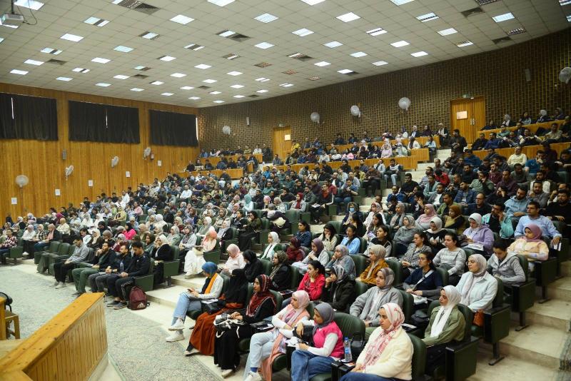
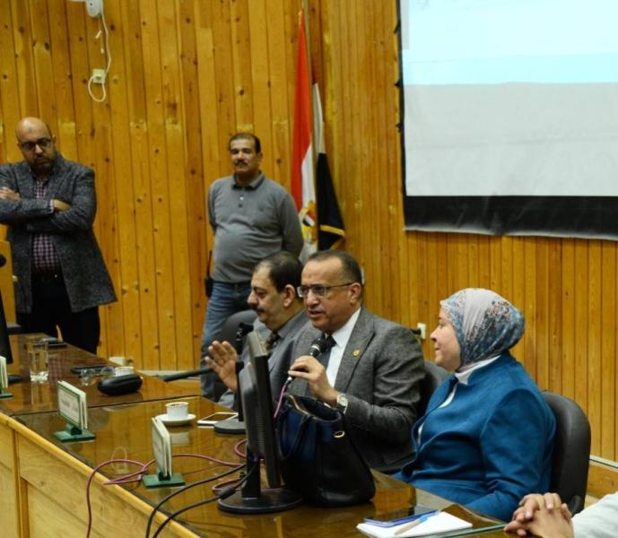
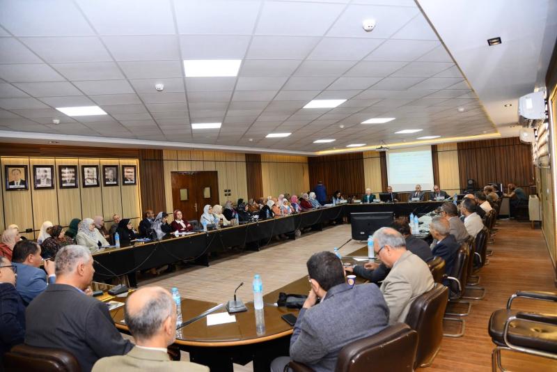
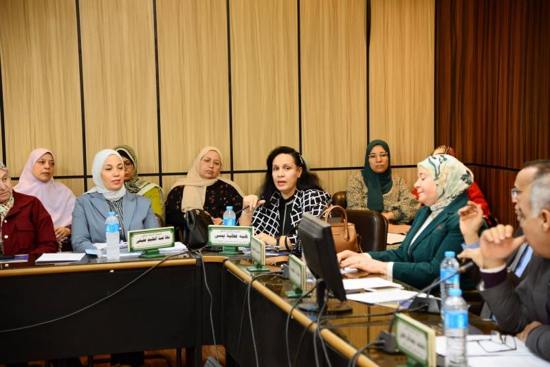
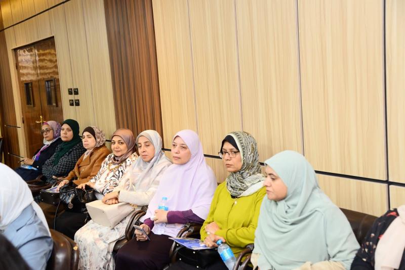
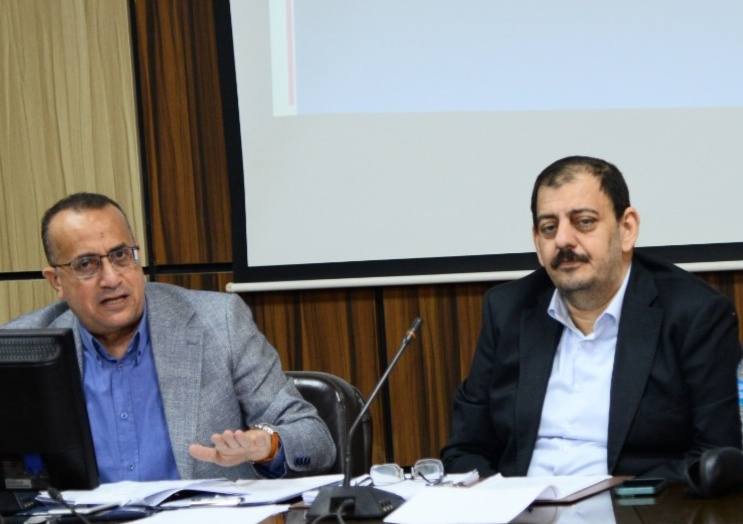
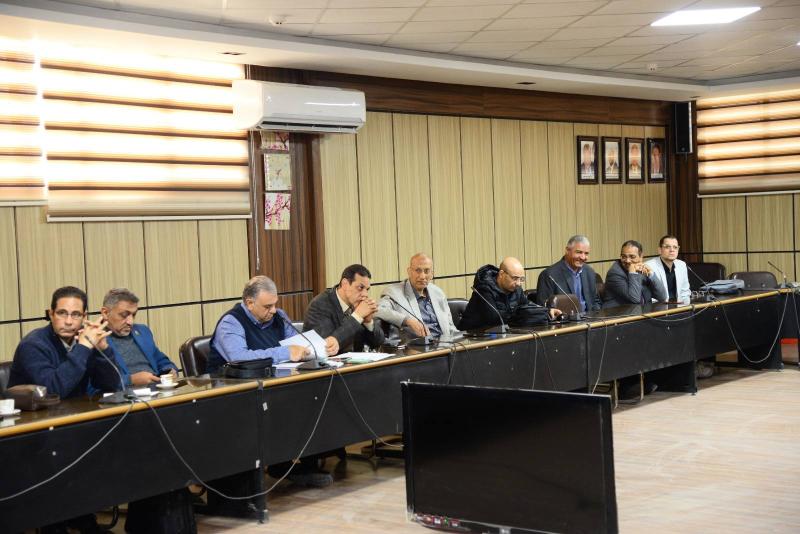
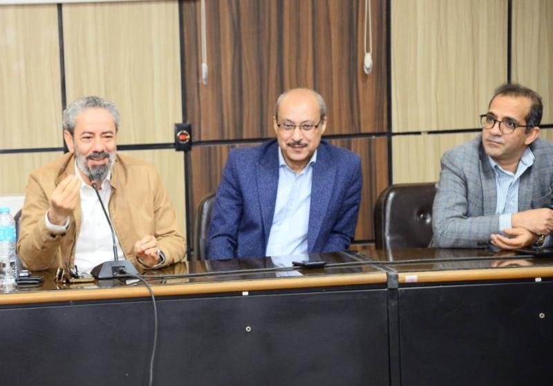
عقد مجلس إدارة مستشفيات جامعة أسيوط اجتماعه الدوري، برئاسة الأستاذ الدكتور علاء عطية، عميد كلية الطب ورئيس مجلس إدارة المستشفيات الجامعية، وبحضور الأستاذ الدكتور خالد عبد العزيز، مدير المستشفى الرئيسي، والأستاذ طارق نجيب سري، رئيس الإدارة المركزية لشئون الأمانة العامة بالمستشفيات الجامعية، والدكتورة إحسان علي حسن رئيس الإدارة المركزية للشئون الطبية والعلاجية، والأستاذة نبيلة أحمد، مدير عام التمريض بالمستشفيات الجامعية، ومديري المستشفيات الجامعية، وذلك لمتابعة مؤشرات الأداء وخطط التطوير بمختلف القطاعات الطبية.
وقد شهد الاجتماع حضور الدكتور محمد جمال، وكيل وزارة الصحة بأسيوط.
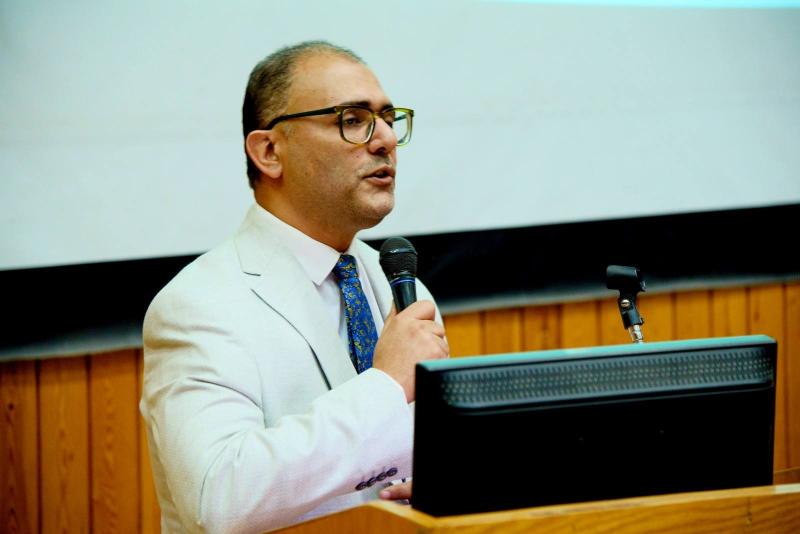
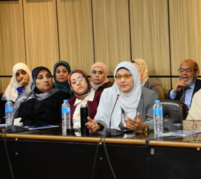
استهل الأستاذ الدكتور علاء عطية أعمال المجلس بتقديم أسمى التهاني للسادة الأعضاء بمناسبة عيد الفطر المبارك، وعقب ذلك، استعرض المجلس التقارير الدورية المتعلقة بمؤشرات الأداء وكفاءة الخدمات الطبية بمختلف المستشفيات الجامعية، حيث شدد على ضرورة تذليل كافة العقبات، بما يضمن تقديم رعاية صحية متكاملة للمرضى.
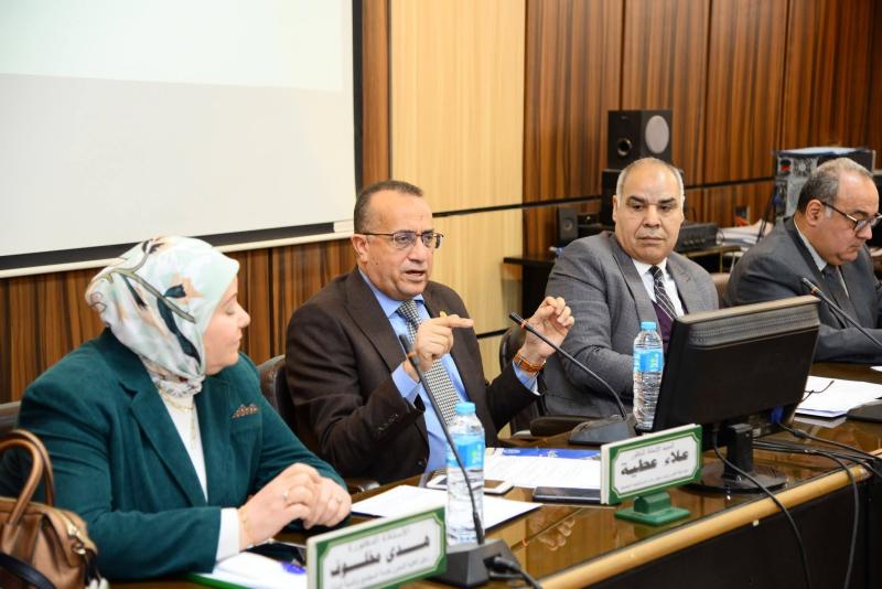
وخلال الاجتماع وجه الأستاذ الدكتور علاء عطية، بمتابعة خطة الطوارئ والأزمات بالمستشفيات، والتحقق من جاهزيتها الكاملة للتعامل مع أي تداعيات طارئة قد تنجم عن تقلبات حالة الطقس،
مؤكداً على أهمية الجاهزية الاستباقية لمواجهة أي ظروف استثنائية ناتجة عن سوء الأحوال الجوية، ضماناً لتقديم الخدمات الطبية اللازمة بكفاءة.
وأكد الأستاذ الدكتور علاء عطية، على أهمية استمرار البرامج التدريبية للأطقم الطبية والتمريضية لرفع كفاءة التعامل مع الحالات الحرجة والعمليات الدقيقة، مؤكدا على أن هذه الخطوات تأتي انطلاقا من الدور الريادي لمستشفيات جامعة أسيوط، باعتبارها الركيزة الأساسية والوجهة الأولى لتقديم الخدمات الطبية المتكاملة في صعيد مصر.
وفي ختام الجلسة، وجه الأستاذ الدكتور علاء عطية، الشكر لكافة الأطقم الطبية والإدارية على جهودهم الملموسة، مشدداً على ضرورة الالتزام بالمعايير القياسية للجودة ومكافحة العدوى في كافة الوحدات العلاجية.
يأتي انعقاد هذا المجلس تأكيدا على إلتزام مستشفيات جامعة أسيوط بالمتابعة الدورية بهدف تقييم الأداء والتأكد من انتظام تقديم كافة الخدمات الطبية في مختلف الأقسام.
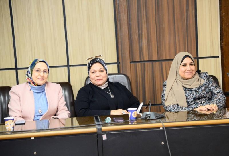
جاء الاجتماع بتنظيم من الأستاذة وفاء عبد السلام محمد، مدير إدارة أمانة مجلس المستشفيات الجامعية.
 Do you have any questions?