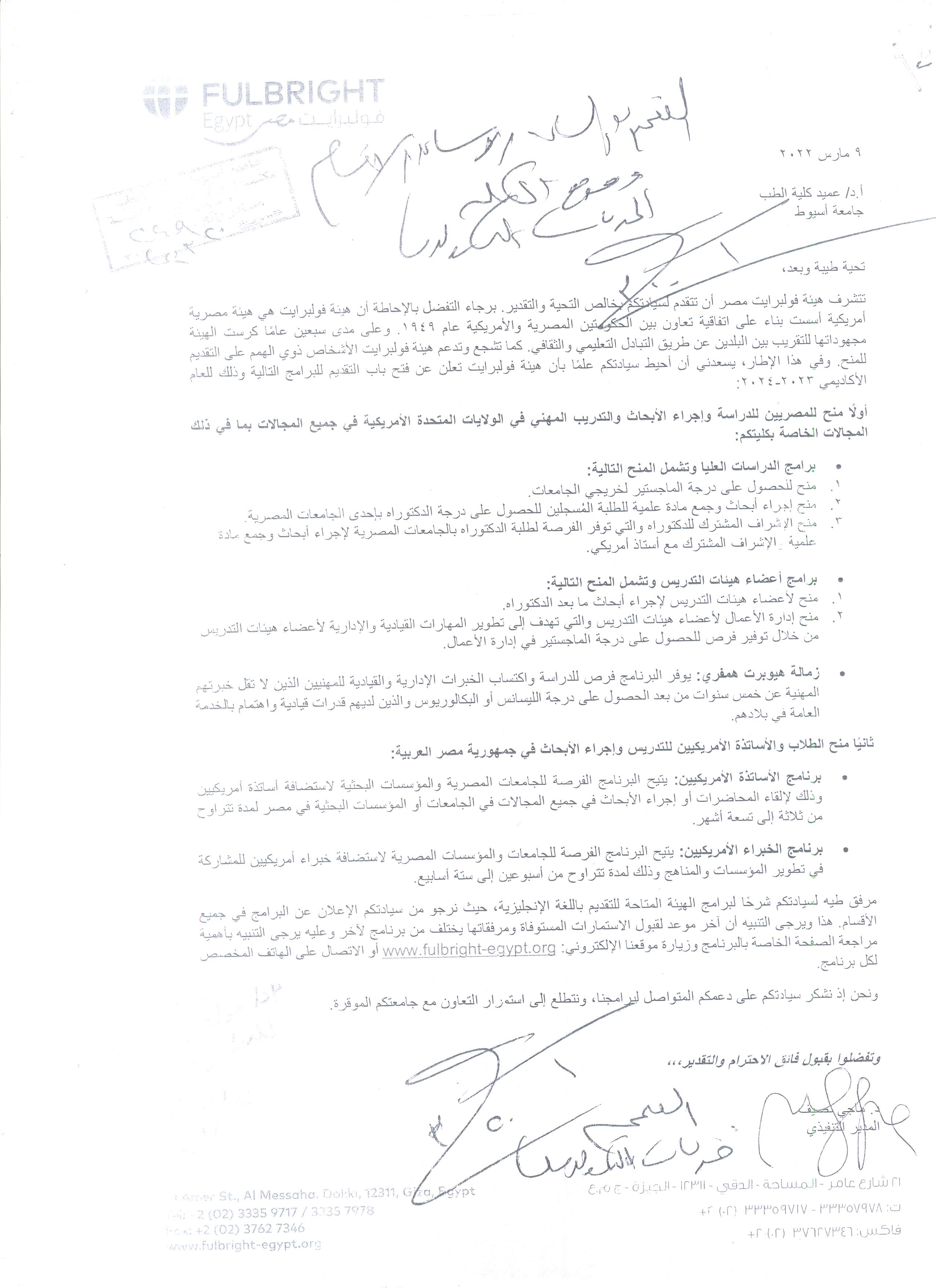
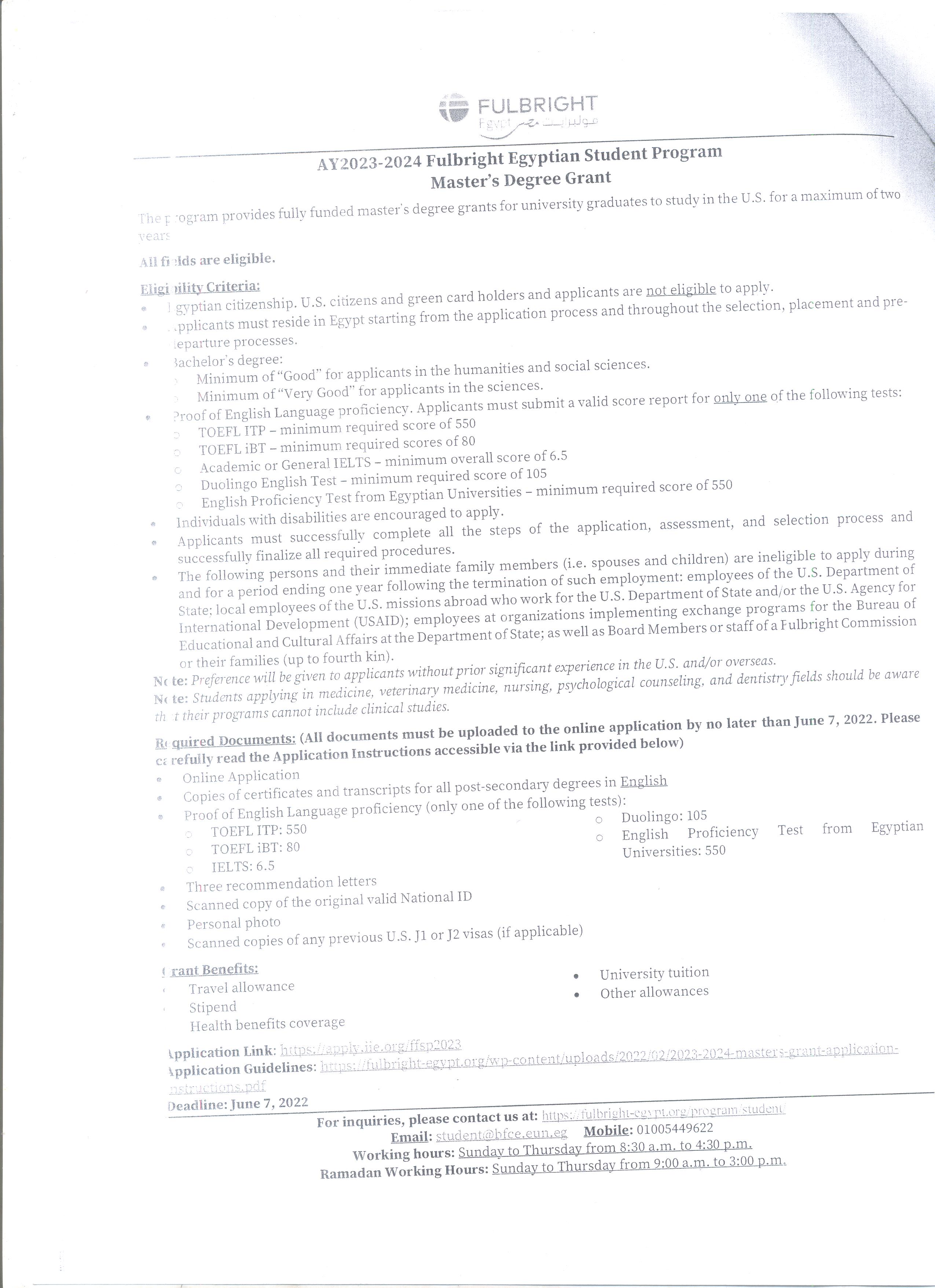
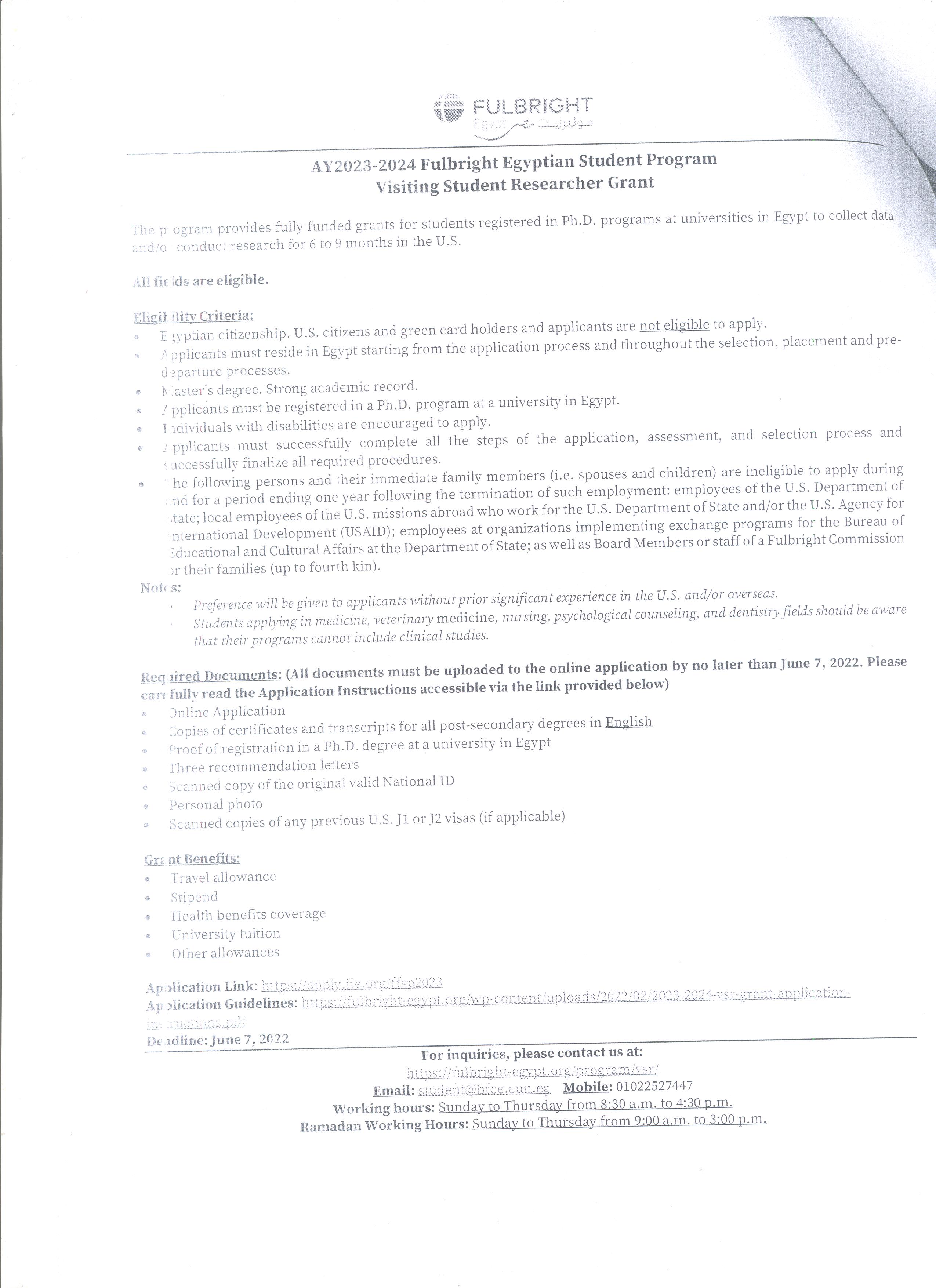
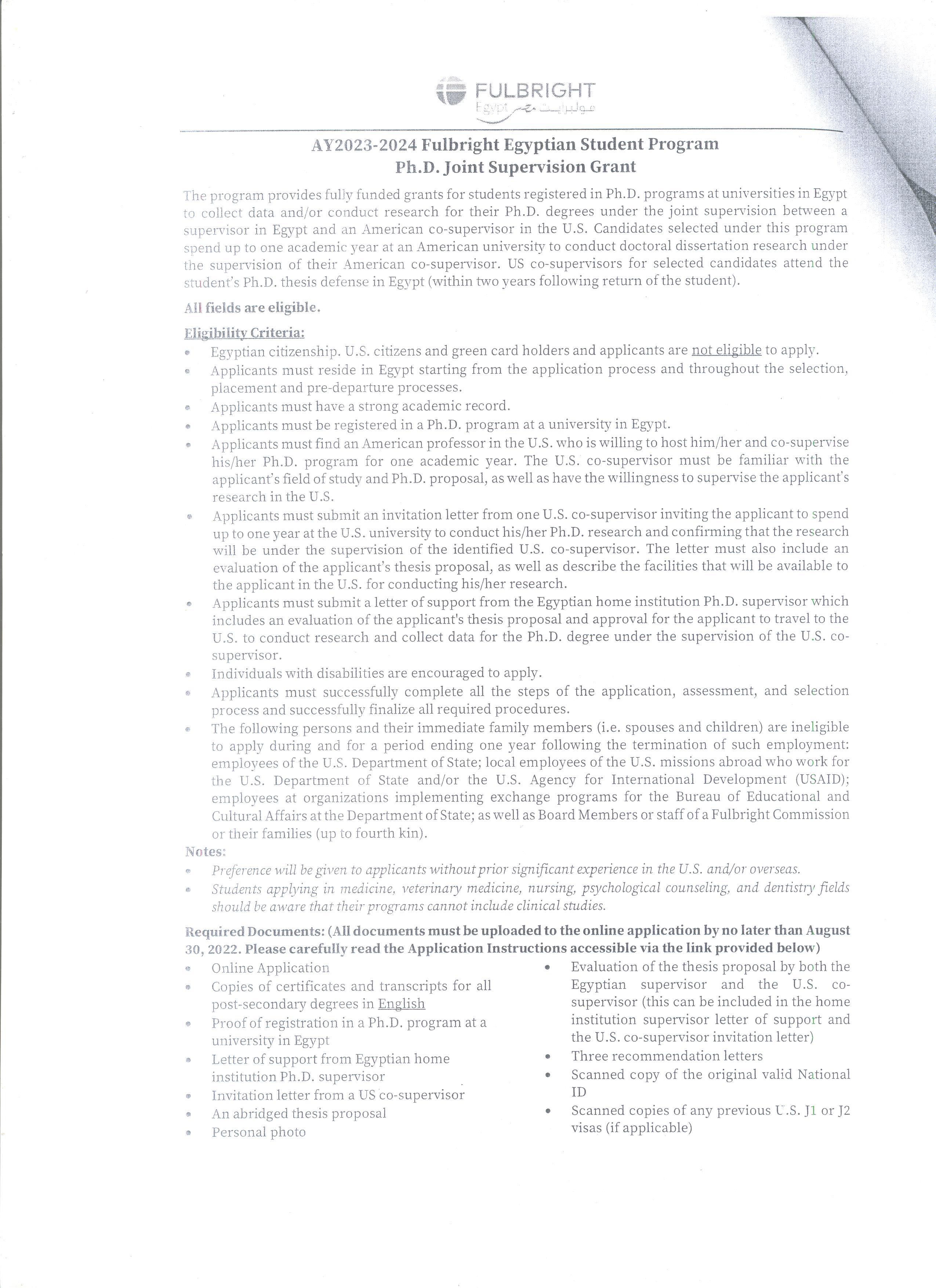
Do you have any questions?
Relationship between noninvasive central blood pressure and brain natriuretic peptide levels in patients with hypertensive pulmonary edema
Research Abstract
Objective
Hypertensive pulmonary edema is a fatal condition unless early and properly diagnosed and managed. Central blood pressure (cBP) has been proven to be more associated with adverse cardiovascular events. We aimed to study the correlation between cBP and heart damage in patients with Hypertensive pulmonary edema.
Methods
We included 50 patients admitted to the emergency department in a university hospital for hypertensive pulmonary edema, 27 women and 23 men aged 50 to 70 years. We excluded patients with suspected acute coronary syndrome, significant valvular heart disease, and pericardial diseases. We measured cBP non-invasively from pulse wave analysis of the brachial artery. Brain natriuretic peptide (BNP) and cBP were repeatedly measured for every patient.
Results
The median BNP levels of patients significantly decreased from 284 pg/ml (232–352.5) to 31.5 pg/ml (24–54) on discharge, P < 0.001. We found a significant correlation between admission BNP and central SBP (cSBP), urea, creatinine, arterial blood gases parameters, and left ventricular end-diastolic diameter (LVEDD). Concurrently, BNP at discharge was correlated with age, central DBP (cDBP), urea, creatinine, LVEDD, partial oxygen pressure (pO2), and oxygen saturation (SO2). Delta BNP was correlated with cSBP, peripheral SBP, urea, creatinine, pO2, and SO2. Linear regression analysis revealed that creatinine, and cSBP, were independent predictors of admission BNP, while urea and cDBP were the independent predictors of discharge BNP.
Conclusion
This simple, noninvasive method of cBP measurement was significantly associated with the extent of myocardial damage in patients presenting with hypertensive pulmonary edema.
Research Date
Research Department
Research Journal
Blood Pressure Monitoring
Research Member
Research Pages
113-120
Research Publisher
Ovid
Research Rank
Q4
Research Vol
2022 Apr 1;27(2)
Research Website
Blood Press Monit. 2022 Apr 1;27(2):113-120. doi: 10.
Research Year
2022
The role of diffusion-weighted imaging MRI in differentiation between benign and malignant axillary lymphadenopathy in clinically TNM stages I and II breast cancer
Research Department
Research Journal
Journal of Current Medical Research and Practice
Research Member
Research Pages
257
Research Publisher
Medknow Publications
Research Vol
6
Research Website
3
Research Year
2021
Radio‑carpal wrist MR arthrography: comparison of ultrasound with fuoroscopy and palpation‑guided injections
Research Department
Research Journal
Skeletal Radiology
Research Member
Research Pages
765 - 775
Research Publisher
Springer
Research Vol
51
Research Year
2021
Association of follicular helper T and follicular regulatory T cells with severity and hyperglycemia in hospitalized COVID-19 patients
Research Abstract
We aimed to determine the levels of follicular helper T (Tfh) and follicular regulatory T (Tfr) cells in COVID-19 patients and determine whether their levels correlated with disease severity and presence of hyperglycemia. This study was carried out in 34 hospitalized COVID-19 patients and 20 healthy controls. Levels of total circulating Tfh, inducible T-cell costimulator (ICOS)+ activated Tfh, and Tfr cells were assessed in all participants by flow cytometry. Total CD4+CXCR5+ Tfh cells and ICOS+Foxp3-activated Tfh cells increased and ICOS+Foxp3+ Tfr cells decreased in COVID-19 patients, especially in diabetic patients and those with severe disease. Activated ICOS+ Tfh cells were directly correlated with lactate dehydrogenase, D-dimer, ferritin, and respiratory rate and inversely correlated with the partial pressure of carbon dioxide. COVID-19 is associated with marked activation of Tfh cells and a profound drop in Tfr cells, especially in severe and diabetic patients. Future studies on expanded cohorts of patients are needed to clarify the relationship between SARS-CoV-2 and acute-onset diabetes.
Research Date
Research Department
Research Journal
Virulence
Research Member
Research Pages
569-577
Research Publisher
Taylor and Francis
Research Vol
13 (1)
Research Website
https://www.tandfonline.com/doi/citedby/10.1080/21505594.2022.2047506?scroll=top&needAccess=true
Research Year
2022