
Announcement of the scientific symposium entitled “Applications of Artificial Intelligence in Digestive and Infectious Diseases”

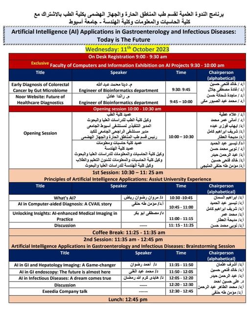
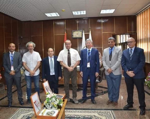
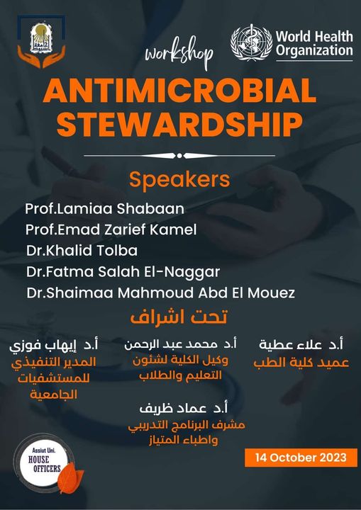
اعلان الندوة العلمية بعنوان " تطبيقات الذكاء الاصطناعي في أمراض الجهاز الهضمي والأمراض المعدية" تحت رعاية الدكتور أحمد المنشاوي -رئيس الجامعة الدكتور علاء عطية- عميد كلية الطب ورئيس مجلس إدارة المستشفيات الجامعية الدكتورة أماني عمر -وكيل الكلية لشئون الدراسات العليا والبحوث الدكتور إيهاب فوزي- المدير التنفيذي للمستشفيات الجامعية الدكتور شريف إبراهيم كامل- مدير مستشفى الراجحي. تأتي الندوة في إطار التعاون بين قسم طب المناطق الحارة والجهاز الهضمي بجامعة أسيوط وكلية الهندسة وكلية الحاسبات والمعلومات. بالتنسيق مع الدكتور نوبي محمد حسن -عميد كلية الهندسة الدكتورة تيسير حسن عبد الحميد- عميد كلية الحاسبات والمعلومات والدكتورة مديحة العطار- رئيس قسم طب المناطق الحارة والجهاز الهضمي. تستهدف الندوة التعرف على كيفية الاستعانة بالتكنولوجيا الحديثة للكشف المبكر عن أمراض الجهاز الهضمي لتجنب حدوث مضاعفات. يأتي اليوم بتنظيم من الدكتورة هايدي كرم الله رمضان، والدكتور أحمد رضوان رياض مقررا الندوة.

The Dean of the College gifts the college shield and the conference shield to the conference guests from the conference

الورشة العلمية لقسم جراحة المسالك البولية من الصين والهند والتي اقيمت في الفترة من  ٨-٩ اكتوبر ٢٠٢٣

استقبل الاستاذ الدكتور علاء عطية عميد كلية الطب الدكتور ضياء عبدالحميد استاذ المسالك ومدير المستشفى والدكتور أحمد الدروي رئيس وحدة جراحة المسالك البولية للأطفال ومقرر الورشة العلمية والدكتور احمد رضا  استاذ مساعد المسالك البولية والدكتور عمرو مهران مدرس المسالك البولية وأعضاء اللجنة المنظمة للورشة العلمية وضيوف الورشة العلمية لقسم جراحة المسالك البولية من الصين والهند والتي اقيمت في الفترة من  ٨-٩ اكتوبر ٢٠٢٣ وهم Prof.Guohua Zeng نائب رئيس جامعة جوانزو ورئيس  الجمعية الدولية لحصوات المسالك البولية بالصين

 و Prof.S.K.Pal رئيس شعبة الحصوات بالجمعية الهندية لجراحة المسالك البولية بجامعة نيودلهي بالهند.

وقام الدكتور علاء عطية بإهدائهم درع الكلية ودرع الورشة العلمية تقديراً وامتنانا على مجهودهم ومشاركتهم المتميزة بورشة العمل.

والورشة تحت إشراف الأستاذ الدكتور محمد أحمد شلبي رئيس شرف الورشة العلمية والاستاذ الدكتور ياسر عبد السلام رئيس القسم والأستاذ الدكتور أحمد الدوري المقرر العام للورشة والدكتور أحمد عبد الحميد شحات الأستاذ المساعد بالقسم  ورئيس اللجنة المنظمة للورشة وقد تناولت الجوانب المختلفة لحصوات الكلى في الأطفال طبياً وجراحياً ، كما شهدت عمليات ذات تقنية عالية تستخدم  لأول مرة في مصر و تم إجراء 17 عملية جراحية على مدار اليومين، كما تم تدريب المشاركين على استخدام منظار الحالب المرن، وإجراء العمليات باستخدام منظار الكلى و منظار الحالب المرن.

كما تم تنسيق ندوة علمية مع أساتذة من أقسام كلى أطفال ، تغذية أطفال ، التخدير ، الباثولوجيا الإكلينيكية والأشعة لمناقشة الجوانب الطبية والعوامل المؤثرة في زيادة تكوين الحصوات لدى الأطفال وكيفية الوقاية منها ومناقشة سبل التشخيص والعناية بالأطفال قبل العملية وأثناءها وبعدها.

وقد أشاد الأطباء الأجانب بمدى جاهزية غرف العمليات والتي تقترب من مستشفيات أوروبا حسب تعبيرهم ومستوي الجراحين العالي بالمستشفى.

والجدير بالذكر أن مستشفى المسالك البولية تعد أكبر مستشفى متخصص في صعيد مصر.

A Comparative Evaluation of Mediastinal Nodal SUVmax and Derived Ratios from 18F-FDG PET/CT Imaging to Predict Nodal Metastases in Non-Small Cell Lung Cancer

Research Abstract

18F-FDG positron emission tomography with computed tomography (PET/CT) is a standard imaging modality for the nodal staging of non-small cell lung cancer (NSCLC). To improve the accuracy of pre-operative staging, we compare the staging accuracy of mediastinal lymph node (LN) standard uptake values (SUV) with four derived SUV ratios based on the SUV values of primary tumours (TR), the mediastinal blood pool (MR), liver (LR), and nodal size (SR). In 2015–2017, 53 patients (29 women and 24 men, mean age 67.4 years, range 53–87) receiving surgical resection have pre-operative evidence of mediastinal nodal involvement (cN2). Among these, 114 mediastinal nodes are resected and available for correlative PET/CT analysis. cN2 status accuracy is low, with only 32.5% of the cN2 cases confirmed pathologically. Using receiver operating characteristic (ROC) curve analyses, a SUVmax of N2 LN performs well in predicting the presence of N2 disease (AUC, 0.822). Based on the respective selected thresholds for each ROC curve, normalisation of LN SUVmax to that for mediastinum, liver and tumour improved sensitivities of LN SUVmax from 68% to 81.1–89.2% while maintaining acceptable specificity (68–70.1%). In conclusion, normalised SUV ratios (particularly LR) improve current pre-operative staging performance in detecting mediastinal nodal involvement.

Research Authors
Maha AlRasheedi, Sai Han, Helene Thygesen, Matt Neilson, Fraser Hendry, Ahmed Alkarn, John D. Maclay, Hing Y. Leung
Research Date
Research Journal
Diagnostics
Research Member
Research Vol
13

Diagnostic Role of Grayscale and Color Doppler Transthoracic Ultrasonography in Pulmonary Thromboembolism in the Emergency Room

Research Abstract

PURPOSE: Pulmonary thromboembolism (PTE) remains under-diagnosed fatal disease at the emergency units suggesting the need for alternative, easy, noninvasive bedside diagnostic approaches. The aim of this study was to determine the diagnostic role of transthoracic ultrasonography (TUS) in PTE, and to evaluate if there is any additive diagnostic value of Color Doppler over gray-scale TUS.

METHODS: Blinded to 64 multi-detectors CT pulmonary angiography (MDCTPA) examination as a gold standard, 60 patients with clinically suspected PTE underwent gray-scale and Color Doppler TUS examination. Results were compared and diagnostic accuracy of TUS was assessed

RESULTS: In 33 patients out of 40 MDCTPA proved cases of PTE, TUS demonstrated 66 lesions with mean (SD) 2±1.20 lesions /patient. For Gray-scale TUS, sensitivity, specificity, positive and negative predictive values and accuracy were 82%, 90%, 94%, 72% and 85%, meanwhile color Doppler TUS had values of 80%, 95%, 97%, 70.4% and 87.5%, respectively.

CONCLUSIONS: TUS is a reliable screening technique for diagnosing PTE with high specificity but relatively low sensitivity. Adding Color Doppler to Gray-scale TUS increases the specificity and consequently the confidence in the diagnosis of peripheral pulmonary infarctions and differentiates them from other pulmonary lesions.

CLINICAL IMPLICATIONS: TUS, in trained hands, has high specificity and diagnostic accuracy allowing its use as bedside screening method in emergency-based situations, especially for critically ill and immobile patients that allows initiation of anticoagulants.

Research Authors
Maha Ghanem, Hoda Makhlouf, Ali Abd ElAzim, Ahmed Al Karn
Research Date
Research Journal
Pulmonary Vascular Disease
Research Member
Research Vol
148

Role Of Bedside Chest Ultrasonography And Echocardiography In The Diagnosis Of Pulmonary Embolism In Emergency Rooms

Research Authors
Maha K. Ghanem , Houda Makhlouf , Ali Abd El-Azim , Ahmed A. AlKarn
Research Date
Research Journal
American Journal of Respiratory and Critical Care Medicine
Research Member
Research Vol
193

C65 ONE STEP UP FROM PE TO CTEPH: Role Of Bedside Chest Ultrasonography And Echocardiography In The Diagnosis Of Pulmonary Embolism In Emergency Rooms

Research Abstract

Introduction: Pulmonary embolism (PE) remains under-diagnosed fatal disease at the emergency units suggesting the need for alternative, easy, noninvasive bedside diagnostic approaches. Objectives: The aim of this study was to determine the diagnostic role of transthoracic ultrasonography (TUS) and echocardiography in PE. Also, to evaluate if there is any additive diagnostic value of Color Doppler over gray-scale TUS in PE. Methods: Blinded to 64 multi-detectors CT pulmonary angiography (MDCTPA) examination as a gold standard, 60 patients with clinically suspected PE underwent gray-scale and Color Doppler TUS examination in addition to echocardiography. Results were compared and diagnostic accuracy of different techniques were assessed. Results:According to Modified Wells Score, 42 (70%) of the cases were defined as moderate risk for PE and 18 (30%) were defined as high risk. In 33 patients out of 40 MDCTPA proved cases of PE, TUS demonstrated 66 lesions with mean (SD) 2±1.20 lesion /patient."Consolidation with little perfusion sign" as detected by colour doppler TUS is helpful in differentiating PE from other peripheral lesions. Using echocardiography, signs of acute right ventricular strain with negative TUS and postive MDCTPA was found in 6 out 7 patients. For Gray-scale TUS, sensitivity, specificity, positive and negative predictive values and accuracy were 82%, 90%, 94%, 72% and 85%, meanwhile color Doppler TUS had values of 80%, 95%, 97%, 70.4% and 87.5%, respectively.. Conclusions: TUS combined with echocardiography is a reliable screening technique for diagnosing PE with high specificity and fair sensitivity. Adding Color Doppler to Gray-scale TUS increases the specificity and consequently the confidence in the diagnosis of peripheral pulmonary infarctions and differentiates them from other pulmonary lesions allowing its use as bedside screening technique in emergency-based situations especially for critically ill and immobile patients that allows initiation of anticoagulants

Research Authors
Ghanem, M K; Makhlouf, H; El-Azim, A Abd; AlKarn, A A Preview author details . 
Research Date
Research Journal
American Journal of Respiratory and Critical Care Medicine
Research Member
Research Vol
193

Factors Affecting Pre-Operative Staging Accuracy in Non-Small Cell Lung Cancer and its Relationship with Survival

Research Abstract

Objectives: Precise staging of non-small cell lung cancer (NSCLC) determines initial treatment and provides more accurate prognostic information for patients. The aim of this cohort study was to determine factors affecting pre- and post-operative mediastinal nodal staging agreement and its effect on 2-year survival.

Materials and Methods: A retrospective multi-centre cohort study was performed, using prospectively collected and pre-defined data from weekly lung cancer multidisciplinary team meetings in 11 hospitals.  Consecutive patients who underwent surgical resection of NSCLC between 2015 and 2017 were eligible. Factors associated with concordant and discordant pre- and post-operative nodal staging, and subsequent lung cancer-specific 2-year mortality were identified by univariate and multivariate regression.

Results: 973 patients fulfilled the eligibility criteria. Concordant pre- and post-operative nodal staging was observed in 783/973 (80%). 123/973 (13%) were under-staged pre-operatively. 67/973 (7%) were over-staged.In 173 patients with clinical N1 or N2 disease (in whom invasive mediastinal staging was indicated), staging EBUS was performed in 55/173 (32%). In these patients, younger age and use of staging EBUS were independent predictors of concordant pre- and post-operative staging.In all patients pre-operative under-staging was independently associated with increased lung cancer-specific 2-year mortality.

Conclusion: Invasive mediastinal staging with EBUS was independently associated with more accurate pre-operative staging. Pre-operative nodal under-staging was associated with increased lung cancer specific mortality. Nodal staging accuracy in potentially curable NSCLC is of fundamental importance to ensure patients receive the correct first-line treatment and to improve survival.

Research Authors
Ahmed Alkarn, Liam Stapleton, Dimitra Eleftheriou, Laura Stewart, George Chalmers, Ahmad Hamed, Khaled Hussein, Kevin G Blyth, Joris van der Horst, John David Maclay
Research Date
Research File
SSRN-id4116418.pdf (267.64 KB)
Research Journal
SSRN
Research Member

Surveillance imaging and recurrence rates following surgery for non-small cell lung cancer (NSCLC)

Research Abstract

Introduction: The evidence base for follow up of NSCLC following surgery remains limited, with variation between guidelines and in clinical practice.

Aims: Review follow up practice, recurrence and subsequent treatment across 11 centres within the West of Scotland.

Methods: Data for 963 consecutive patients treated from 2015-17 were collected prospectively with follow up practice and recurrence within 2 years examined using electronic case notes.

Results: 55% of patients were followed up with plain film, 39% had planned CT scanning with a small proportion lost to follow up.

Overall 26% of patients recurred and this increased with stage (Table 1). Method of recurrence detection is shown in Table 2. 68% of patients had distant metastatic disease at the time of their recurrence diagnosis.

Conclusion: There is significant variability in follow up practice across the West of Scotland. A high proportion of patients who recurred had distant metastatic disease at the time of diagnosis with a small proportion suited for radical treatment. Regular post surgical imaging for patients with higher risk of recurrence may allow more patients to have further radical treatment.

Research Authors
Liam Stapleton, Ahmed Alkarn, John Maclay, Joris Van Der Horst
Research Date
Research Journal
European Respiratory Journal
Research Member
Research Vol
56
Subscribe to